Jonathan A Clare, M.D.
- Assistant Professor of Emergency Medicine

https://www.hopkinsmedicine.org/profiles/results/directory/profile/10004261/jonathan-clare
Zebeta dosages: 10 mg, 5 mg
Zebeta packs: 60 pills, 90 pills, 120 pills, 180 pills, 270 pills, 360 pills
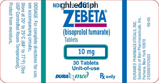
10 mg zebeta purchase fast delivery
The ureter travels within the lateral pelvic wall before coursing medially toward the bladder. In the female pelvis, the ureter travels beneath the broad ligament, lateral to the cervix and under the uterine artery. The ureters join the bladder and journey submucosally within the bladder wall for two to three cm earlier than opening into the bladder at the ureteral orifices. The diagonal course of the intramural section of the ureter helps stop urinary reflux. The ureter consists of two muscle layers, an inside longitudinal layer and an outer round layer. In the wall of the distal ureter, close to its insertion into the bladder, a 3rd muscle layer is continuous with the detrusor muscle of the bladder. The ureter is surrounded by an outer fibrous layer, the adventitia, which is continuous with the renal capsule and the adventitia of the bladder. The belly portions of the ureters obtain their blood provide from a ureteral branch of the renal artery and from branches arising from the aorta, retroperitoneal vessels, gonadal vessels, and iliac vessels. In the abdomen, the arterial branches supplying the ureter are located medial to the ureter, whereas in the pelvis the ureteral arteries arise lateral to the ureter from the iliac, gluteal, obturator, rectal, and vesical arteries. The 826 supplying artery and draining veins travel in the connective tissue surrounding the ureter called the mesoureter. In most sufferers, an anastomosing plexus types within the adventitia along the course of the ureter. The veins of the ureter drain into the renal vein, gonadal vein, lumbar veins, iliac veins, and vesical veins. The lymphatics of the proximal ureter drain into the renal lymphatics, and those of the midureter drain into periaortic and customary iliac lymph nodes. The lymphatics of the distal ureter drain into iliac and presacral lymph nodes and be part of with the lymphatics of the bladder. The innervation of the ureter consists of sympathetic nerves from the aortic plexus and superior and inferior hypogastric plexus that course with the arterial provide to the connective tissue around the ureter. Pelvic splanchnic nerves from the sacral roots present parasympathetic innervation to pelvic viscera. Intrinsic processes might obstruct the lumen, incite irritation and edema in the wall of the ureter, or infiltrate the wall of the ureter. Extrinsic processes could trigger narrowing of the ureter by compression, encasement, or infiltration. Intraluminal filling defects are sometimes utterly surrounded in contrast material. Mucosal and submucosal filling defects are intimately associated with the wall of the ureter and can be differentiated by analysis of the relationship of the lesion to the adjoining ureteral wall, with mucosal lesions typically demonstrating acute angles and submucosal lesions demonstrating obtuse angles. An infiltrative process normally causes an abrupt change in caliber of the ureter with an apple core�like appearance of the concerned ureter. The narrowed segment of ureter demonstrates circumferential wall thickening and mucosal irregularity. This look is classically produced by neoplastic infiltration, however benign causes, such as irradiation, stone disease, and iatrogenic damage, may cause a similar appearance. Encasement of the ureter typically results in a gradual tapered contour and clean mucosal surface. Alternatively, encasement of the ureter could lead to a focal abrupt transition with a dilated proximal ureter and a slender or normal-caliber ureter distally. This appearance on intravenous pyelography has been Document t�l�charg� de ClinicalKey. Many of those processes trigger focal ureteral abnormalities; however, occasionally a multifocal course of happens. Often, other associated imaging findings can help slender the differential analysis, corresponding to the situation of the stricture, focal or multifocal involvement, deviation of the ureter, and involvement of the kidney, bladder, or different organ systems. Disadvantages of this modality embody restricted utility in sufferers with impaired renal operate and poor soft tissue distinction.
Best 5 mg zebeta
Viengchareun S, et al: the mineralocorticoid receptor: insights into its molecular and (patho)physiological biology. Liu W, et al: Steroid receptor heterodimerization demonstrated in vitro and in vivo. Kaspar F, et al: A mutant androgen receptor from sufferers with Reifenstein syndrome: identification of the perform of a conserved alanine residue in the D box of steroid receptors. Sartorato P, et al: Different inactivating mutations of the mineralocorticoid receptor in fourteen households affected by type I pseudohypoaldosteronism. Li Y, et al: Structural and biochemical mechanisms for the specificity of hormone binding and coactivator assembly by mineralocorticoid receptor. Kunzelmann K, Mall M: Electrolyte transport within the mammalian colon: mechanisms and implications for disease. Naray-Fejes-Toth A, et al: sgk is an aldosterone-induced kinase in the renal amassing duct. Shigaev A, et al: Regulation of sgk by aldosterone and its effects on the epithelial Na(+) channel. Schild L, et al: A mutation within the epithelial sodium channel causing Liddle disease increases channel activity in the Xenopus laevis oocyte expression system. Soundararajan R, et al: A novel position for glucocorticoid-induced leucine zipper protein in epithelial sodium channel-mediated sodium transport. Fagart J, et al: Crystal structure of a mutant mineralocorticoid receptor responsible for hypertension. Metivier R, et al: Estrogen receptor-alpha directs ordered, cyclical, and combinatorial recruitment of cofactors on a natural goal promoter. Yokota K, et al: Coactivation of the N-terminal transactivation of mineralocorticoid receptor by Ubc9. Wang H, et al: Clathrin-mediated endocytosis of the epithelial sodium channel: role of epsin. Malik B, et al: Regulation of epithelial sodium channels by the ubiquitin-proteasome proteolytic pathway. Soundararajan R, et al: Epithelial sodium channel regulated by differential composition of a signaling complicated. Diezi J, et al: Micropuncture study of electrolyte transport across papillary collecting duct of the rat. Le Moellic C, et al: Aldosterone and tight junctions: modulation of claudin-4 phosphorylation in renal collecting duct cells. Vandewalle A, et al: Aldosterone binding along the rabbit nephron: an autoradiographic examine on isolated tubules. Gnionsahe A, et al: Aldosterone binding websites along nephron of Xenopus and rabbit. Nishiyama A, et al: Involvement of aldosterone and mineralocorticoid receptors in rat mesangial cell proliferation and deformability. Hierholzer K, Stolte H: the proximal and distal tubular action of adrenal steroids on Na reabsorption. Oberleithner H, et al: Aldosterone prompts Na+/H+ change and raises cytoplasmic pH in target cells of the amphibian kidney. Vallet M, et al: Pendrin regulation in mouse kidney primarily is chloride-dependent. Loffing J, et al: Localization of epithelial sodium channel and aquaporin-2 in rabbit kidney cortex. Clauss W, et al: Ion transport and electrophysiology of the early proximal colon of rabbit. Asher C, et al: Aldosterone-induced improve within the abundance of Na+ channel subunits. Saudan P, et al: Safety of low-dose spironolactone administration in persistent haemodialysis patients. Gross E, et al: Effect of spironolactone on blood strain and the renin-angiotensin-aldosterone system in oligo-anuric hemodialysis sufferers.
Diseases
- Benign mucosal pemphigoid
- Cataract mental retardation hypogonadism
- Epidermodysplasia verruciformis
- Brachymesophalangy mesomelic short limbs osseous anomalies
- Chromosome 2, monosomy 2p22
- Acute megakaryoblastic leukemia
- Cytomegalic inclusion disease
- Birdshot chorioretinopathy
Generic 5 mg zebeta otc
Deposits are bilateral and symmetric and occur subepithelially within the lamina propria of the seminal vesicles in aggregates varying in size from microscopic to grossly seen seminal vesicle wall thickening. When the amyloid deposits are due to systemic amyloidosis, the amyloid is located in the partitions of blood vessels or inside muscle somewhat than in a subepithelial location. Amyloid is demonstrated as areas of low echogenicity which may be indistinguishable from different seminal vesicle masses. Biopsy helps affirm the diagnosis and rule out local invasion from tumors of adjacent organs. Hoshi A, Nakamura E, Higashi S, et al: Epithelial stromal tumor of the seminal vesicle. Lawrentschuk N, Pan D, Stillwell R, et al: Implications of amyloidosis on prostatic biopsy. The causes of impotence could be psychogenic, endocrinologic, neurogenic, anatomic, infectious, pharmacologic, or vasogenic. Relaxation of the smooth muscles of the cavernous and helicine arteries causes their dilatation. Venous incompetence or failure of the venoocclusive mechanism of outflow restriction has been recognized as a significant factor and may be its most typical trigger. Perineal radiation therapy may affect the frequent penile artery near the prostate gland. Prolonged high-flow Prevalence and Epidemiology With rising societal openness toward sexual dysfunction, more males are coming ahead for evaluation and administration of erectile dysfunction. Pathophysiology Penile erection is a neurovascular phenomenon ensuing from arterial dilatation, sinusoidal leisure, and venous outflow restriction. Arterial compliance and sufficient blood inflow are important to achieve erection, and restriction of venous outflow is essential to preserve it. However, this index could also be affected by aortoiliac disease, even when penile hemodynamics are regular. At present, penile Doppler ultrasonography is the mainstay of imaging analysis of impotence. Warm saline and low osmolar contrast material is infused via one corpus cavernosum, perfusing the opposite through anastomotic connections. Direct pressure measurements are obtained through the needle in the different corpus cavernosum. After baseline measurement of intracavernosal pressure and penile circumference, pharmacologic erection is produced by intracavernosal injection. Pressure and penile circumference monitoring is then carried out for 10 minutes or till equilibrium happens. If no erection is produced, heparinized saline is infused at more and more speedy charges until intracavernosal stress of one hundred fifty mm Hg is achieved. Venous outflow resistance also may be assessed by observing the rate of intracavernosal stress fall after termination of intracavernosal saline infusion. In cavernosography, a hundred to 150 mL of contrast agent is infused into the corpus cavernosum to preserve 90 mm Hg of intracavernosal stress. Fluoroscopy and spot films through the procedure may reveal the site of venous leak and provide the preprocedural anatomic information. Aortoiliac arteriography is carried out to evaluate for proximal atherosclerotic lesions and the patency of the inferior epigastric arteries, which are normally used for surgical revascularization. A high-frequency linear transducer is used with a mechanical standoff wedge to produce a good insonating angle. Optimization of "slow flow" sensitivity is essential for the correct depiction of diastolic flow and blood flow in the dorsal vein. Ultrasound evaluation of the flaccid penis is first performed to assess for structural anomalies and plaques. B, With the onset of erection or after papaverine injection there is a rise in each systolic and diastolic flow in the cavernous artery. A B the normal peak systolic velocity in the erect penis ranges from 35 to 60 cm/s. As the intracavernosal strain will increase, a dicrotic notch seems on the finish of systole accompanied with the lower in diastolic flow. Thus, a high-resistance waveform must be seen within the cavernosal artery at erection, with little or no diastolic move.
Zebeta 5 mg discount on line
This group consists of illnesses causing nephrocalcinosis and those who current primarily with calyceal or papillary abnormalities. Yes No Unilateral small, easy kidney Bilateral small, easy kidneys Lobar infarction Dilated calyces at renal poles Widespread papillary 1. Vascular � Generalized arteriosclerosis � Benign/malignant nephrosclerosis � Atheroembolic renal disease � Arterial hypotension 2. It manifests as nephritic syndrome, with a positive antistreptolysin O titer as diagnostic, and restoration is the rule in 2 to three weeks. It is often idiopathic however may be associated with cancers (lung, bowel), infection (hepatitis, malaria), medicine (penicillamine), and systemic lupus erythematosus. Also observe that the renal contour is easy and the traditional pelvicalyceal relationships are preserved. Uncommonly, there could additionally be replacement of the wasted renal tissue with fatty proliferation within the renal sinus (renal sinus lipomatosis). Diagrammatic representation of the pyelocalyceal and corresponding renal contour abnormalities. A, Reflux nephropathy, which shows papillary abnormality with overlying scar generally seen at the renal poles. C, Papillary necrosis displaying random papillary abnormalities without cortical involvement. There may be hypercellularity with thickening of the glomerular basement membrane, hyalinization with deposition of amorphous proteinaceous materials, and sclerosis that results in obliteration of the glomerular tuft. Emphysematous pyelonephritis is a necrotizing an infection of the renal parenchyma seen primarily in diabetic sufferers. These sufferers typically have comorbid conditions similar to diabetes, being pregnant, systemic illness, or persistent liver illness. Complicated pyelonephritis is associated with recurrent disease, structural abnormalities, diabetes, pregnancy, immunosuppression, and extended signs (>2 weeks in duration). Predisposing elements for pyelonephritis embody urinary tract obstruction, vesicoureteral reflux, pregnancy, urinary tract instrumentation, preexisting renal disease or systemic predisposition similar to diabetes mellitus, and immunosuppression. It is extra common in adults than in youngsters, in females youthful than the forty years of age, and in males older than the age of sixty five years. It is most commonly seen in kids owing to the much larger incidence of vesicoureteral reflux on this inhabitants. It is two to six instances extra frequent in females than in males23 and is often seen in diabetic patients. An abnormal host response leads to destruction and replacement of the renal parenchyma by lipid-laden macrophages. Hypercalcemia � Hyperparathyroidism � Milk-alkali syndrome � Hypervitaminosis D � Sarcoidosis � Prolonged immobilization � Metastatic calcification 2. Acute cortical necrosis Algorithm exhibiting approach to diffuse renal parenchymal ailments with normal renal bulk and contour. The presentation of pyelonephritis within the pediatric age group is completely different from that within the grownup and may be difficult to the clinician. Imaging is therefore of higher consideration even within the presence of uncomplicated illness or single episodes. Laboratory knowledge may present an elevated erythrocyte sedimentation price, elevated white blood cell count, and proteinuria. Emphysematous pyelonephritis might manifest as a crepitant mass, a very suggestive discovering. The laboratory research might present hyperglycemia, acidosis, electrolyte imbalance, and thrombocytopenia. Microscopic examination shows interstitial or tubular necrosis and mononuclear cell infiltrate with fibrosis. Chronic pyelonephritis seems as a small shrunken kidney with scars and blunted calyces.
5 mg zebeta discount visa
Kuhn M: Cardiac and intestinal natriuretic peptides: insights from genetically modified mice. Tavi P, Laine M, Weckstrom M, et al: Cardiac mechanotransduction: from sensing to disease and treatment. Capasso G: A new cross-talk pathway between the renal tubule and its personal glomerulus. Morita H, Matsuda T, Tanaka K, et al: Role of hepatic receptors in controlling physique fluid homeostasis. Morita H, Matsuda T, Furuya F, et al: Hepatorenal reflex plays an essential function in natriuresis after high-NaCl meals consumption in acutely aware canines. Morita H, Ohyama H, Hagiike M, et al: Effects of portal infusion of hypertonic resolution on jejunal electrolyte transport in anesthetized canine. Matsuda T, Morita H, Hosomi H, et al: Response of renal nerve activity to excessive NaCl meals intake in canine with continual bile duct ligation. Morita H, Fujiki N, Hagiike M, et al: Functional proof for involvement of bumetanide-sensitive Na+K+2Cl- cotransport within the hepatoportal Na+ receptor of the Sprague-Dawley rat. Koyama S, Kanai K, Aibiki M, et al: Reflex increase in renal nerve activity during acutely altered portal venous strain. Thomas L, Kumar R: Control of renal solute excretion by enteric alerts and mediators. Kinoshita H, Fujimoto S, Nakazato M, et al: Urine and plasma ranges of uroguanylin and its molecular varieties in renal diseases. Fukae H, Kinoshita H, Fujimoto S, et al: Changes in urinary ranges and renal expression of uroguanylin on low or high salt diets in rats. Oppermann M, Mizel D, Huang G, et al: Macula densa control of renin secretion and preglomerular resistance in mice with selective deletion of the B isoform of the Na,K,2Cl co-transporter. Interaction between renal sympathetic nerves and the renin-angiotensin system in the control of renal operate. Li L, Mizel D, Huang Y, et al: Tubuloglomerular suggestions and renal operate in mice with focused deletion of the sort 1 equilibrative nucleoside transporter. Satriano J, Wead L, Cardus A, et al: Regulation of ecto-5nucleotidase by NaCl and nitric oxide: potential roles in tubuloglomerular feedback and adaptation. Zhang Q, Lin L, Lu Y, et al: Interaction between nitric oxide and superoxide within the macula densa in aldosterone-induced alterations of tubuloglomerular suggestions. Schnermann J: Maintained tubuloglomerular suggestions responses throughout acute inhibition of P2 purinergic receptors in mice. Jin C, Hu C, Polichnowski A, Jr: Effects of renal perfusion pressure on renal medullary hydrogen peroxide and nitric oxide production. Matsushima Y, Akabane S, Ito K: Characterization of alpha 1- and alpha 2-adrenoceptors instantly related to basolateral membranes from rat kidney proximal tubules. Effects of guanethidine on the renal response to sodium deprivation in regular man. Friberg P, Meredith I, Jennings G, Jr: Evidence for increased renal norepinephrine overflow during sodium restriction in humans. Kiuchi-Saishin Y, Gotoh S, Furuse M, et al: Differential expression patterns of claudins, tight junction membrane proteins, in mouse nephron segments. Romano G, Favret G, Damato R, et al: Proximal reabsorption with changing tubular fluid influx in rat nephrons. Inagami T, Mizuno K, Kawamura M, Jr: Localization of components of the renin-angiotensin system within the kidney and sustained release of angiotensins from isolated and perfused kidney. Schindler C, Bramlage P, Kirch W, et al: Role of the vasodilator peptide angiotensin-(1-7) in cardiovascular drug remedy. Bankir L: Antidiuretic action of vasopressin: quantitative aspects and interaction between V1a and V2 receptor-mediated results. Brown D, Hasler U, Nunes P, et al: Phosphorylation occasions and the modulation of aquaporin 2 cell floor expression. Aoyagi T, Izumi Y, Hiroyama M, et al: Vasopressin regulates the renin-angiotensin-aldosterone system via V1a receptors in macula densa cells. Yasuoka Y, Kobayashi M, Sato Y, et al: Decreased expression of aquaporin 2 in the collecting duct of mice missing the vasopressin V1a receptor. Kishimoto I, Tokudome T, Nakao K, et al: Natriuretic peptide system: an overview of studies using genetically engineered animal models.
Zebeta 10 mg cheap fast delivery
Such calcifications are additionally encountered in patients with pseudohypoparathyroidism. Bone disease could also be noticed, however its findings differ within the varied causes of hypocalcemia (see later). These entities should be thought of early in the prognosis of hypocalcemic individuals. A thorough medical history and physical examination are diagnostically necessary as a outcome of hypocalcemia can be brought on by postsurgical, pharmacologic, inherited, developmental, and nutritional issues, along with being a part of advanced syndromes. All these situations present in the course of the neonatal period with extreme hypocalcemia without some other organ involvement and reply properly to therapy with vitamin D analogues. Treatment with calcium supplements and vitamin D is warranted just for patients with severe symptomatic hypocalcemia. The goal should be to enhance the calcium degree to render the patient asymptomatic, not essentially to a normocalcemic degree. Renal calcium excretion requires monitoring as a outcome of these sufferers may develop frank hypercalciuria and nephrocalcinosis. This situation, reported in 1942 by Albright, was the primary described example of a hormone resistance disease. Congenital defects leading to hypomagnesemia and hypocalcemia are mentioned later (see "Magnesium Disorders") and in Chapters forty three and 75. Acquired Hypoparathyroidism and Inadequate Parathyroid Hormone Production Postsurgical Causes. The commonest cause of acquired hypoparathyroidism in adults is surgical removal of or harm to the parathyroid glands. Transient hypocalcemia after thyroid surgery was observed in 2% to 23% of cases, whereas permanent hypocalcemia occurred in roughly 1% to 2%. Hypoparathyroidism could end result from inadvertent removing of the parathyroids, damage from bleeding, or devascularization. Surgical experience and use of appropriate surgical method might scale back the frequency of hypothyroidism. The mixture of calcium deficiency and vitamin D deficiency accelerates skeletal abnormalities and the event of hypocalcemia. A number of completely different mutations have been found within the vitamin D receptor gene of affected individuals. Inherited and acquired disorders of vitamin D and its metabolites may be related to hypocalcemia. Vitamin D is current naturally in a few foods, is artificially added to others, and is on the market as a meals supplement or drug. Vitamin D deficiency with hypocalcemia is commonly seen in patients with renal insufficiency (see Chapter 55). Prolonged vitamin D deficiency causes rickets in kids (a disorder of Medications. Medication-induced hypocalcemia is a comparatively frequent cause of hypocalcemia, notably in hospitalized sufferers. Calcium readings may be as low as 6 mg/dL, however with no symptoms or indicators of hypocalcemia. Foscarnet can cause hypocalcemia through the chelation of extracellular calcium ions, so normal whole calcium measurements could not mirror ionized hypocalcemia. Patients handled with foscarnet should bear total calcium and ionized calcium measurements. Oral sodium phosphate�induced hyperphosphatemia may trigger hypocalcemia, significantly in patients with renal failure. In difficult, critically unwell sufferers, total calcium measurements could also be poor indicators of the ionized calcium focus because many components that might intrude with or alter calcium and protein binding could additionally be current. It is probably because of calcium chelation by free fatty acids generated by the motion of pancreatic lipase, although some animal studies have challenged this speculation. The therapy is determined by pace of onset and the severity of scientific and laboratory features.
Agarweed (Agar). Zebeta.
- Are there safety concerns?
- Are there any interactions with medications?
- Constipation, diabetes, weight loss, and obesity.
- How does Agar work?
- What is Agar?
- Dosing considerations for Agar.
Source: http://www.rxlist.com/script/main/art.asp?articlekey=96124
Buy zebeta 10 mg fast delivery
The white substance masking his head is vernix caseosa, a traditional fatty protective overlaying. The iris acquires its definitive color as pigmentation occurs in the course of the first 6 to 10 months. The focus and distribution of pigment-containing cells (chromatophores) in the free vascular connective tissue of the iris decide eye shade. If the melanin pigment is confined to the pigmented epithelium on the posterior floor of the iris, the iris appears blue. If melanin can be distributed throughout the stroma (supporting tissue) of the iris, the attention seems brown. Iris heterochromia also can outcome from changes to the sympathetic innervations to the attention. The inner layer of the optic cup has thickened to type the primordial neural retina. The outer layer is heavily pigmented and is the primordium of the pigment layer of the retina. Cardiac defects and deafness are other delivery defects generally attributed to this infection. Aniridia could also be familial (occurring in members of a family); the trait could also be transmitted in a dominant or sporadic sample. These cells lengthen significantly to type extremely clear epithelial cells, the first lens fibers. Although secondary lens fibers proceed to form throughout adulthood and the lens will increase in diameter, the first lens fibers must final a lifetime. However, it becomes avascular in the fetal interval, when this part of the hyaloid artery degenerates. The pupillary membrane develops from the mesenchyme posterior to the cornea in continuity with the mesenchyme growing within the sclera. This capsule represents a tremendously thickened basement membrane and has a lamellar structure due to its development. It consists of vitreous humor, which is the fluid part of the vitreous physique. The main vitreous humor is derived from mesenchymal cells of neural crest origin, which secrete a gelatinous matrix; this surrounding substance is recognized as the primary vitreous body. The major humor is surrounded later by a gelatinous secondary vitreous humor, which is assumed to arise from the inner layer of the optic cup. The secondary humor consists of primitive hyalocytes (vitreous cells), collagenous materials, and traces of hyaluronic acid. The hyaloid artery remnant generally might seem as a fantastic strand traversing the vitreous physique. In unusual circumstances, the complete distal part of the artery persists and extends from the optic disc by way of the vitreous body to the lens. The mesenchyme superficial to this area types the substantia propria (transparent connective tissue) of the cornea and the mesothelium of the anterior chamber. After the lens is established, it induces the floor ectoderm to become the epithelium of the cornea and conjunctiva. The posterior chamber of the eye develops from a space that varieties within the mesenchyme posterior to the creating iris and anterior to the developing lens. Intraocular rigidity rises because of an imbalance between the production of aqueous humor and its outflow. Congenital glaucoma is genetically heterogeneous (includes several phenotypes that appear comparable however are decided by completely different genotypes), however the condition may also result from a rubella an infection during early being pregnant (see Chapter 20, Table 20-6). Rarely, the entire pupillary membrane persists, giving rise to congenital atresia of pupil (absence of a pupil opening). This vascular structure encircling the anterior chamber of the eye is the outflow website of aqueous humor from the anterior chamber to the venous system. The sclera develops from a condensation of mesenchyme external to the choroid and is continuous with the stroma (supporting tissue) of the cornea. The first choroidal blood vessels seem through the 15th week; by the 23rd week, arteries and veins may be easily distinguished.
Buy cheap zebeta 5 mg on line
Conversion of Phylloquinone (Vitamin K1) into Menaquinone-4 (Vitamin K2) in Mice two attainable routes for menaquinone-4 accumulation in cerebra of mice. The manufacturing of menaquinones (vitamin K2) by intestinal micro organism and their function in sustaining coagulation homeostasis. Proteins involved in uptake, intracellular transport and basolateral secretion of fat-soluble vitamins and carotenoids by mammalian enterocytes. Hypercoagulable state and thromboembolism following warfarin withdrawal in post-myocardial-infarction sufferers. Initiation of warfarin in sufferers with atrial fibrillation: Early effects on ischaemic strokes. Vitamin K diet, metabolism, and require ments: Current ideas and future research. The propeptide of rat bone gamma-carboxyglutamic acid protein shares homology with other vitamin K-dependent protein precursors. The propeptides of the vitamin K-dependent proteins possess totally different affinities for the vitamin K-dependent carboxylase. Vitamin K-dependent gammacarbon-hydrogen bond cleavage and nonmandatory concurrent carboxylation of peptide-bound glutamic acid residues. Structural and practical insights into human vitamin K epoxide reductase and vitamin K epoxide reductase-like1. Linkages between genes for coat colour and resistance to warfarin in Rattus norvegicus. Homozygosity mapping of a second gene locus for hereditary mixed deficiency of vitamin K� dependent clotting factors to the centromeric area of chromosome 16. Engineering of a recombinant vitamin Kdependent -carboxylation system with enhanced -carboxyglutamic acid forming capability proof for a functional cxxc redox center within the system. The conversion of vitamin K epoxide to vitamin K quinone and vitamin K quinone to vitamin K hydroquinone uses the identical active website cysteines. Human vitamin K epoxide reductase and its bacterial homologue have different membrane topologies and reaction mechanisms. The synthesis of the -diketone derived from the hemorrhagic agent via alkaline degradation. Warfarin binding to microsomes isolated from regular and warfarin-resistant rat liver. Pharmacoki netic interaction between warfarin and a uricosuric agent, bucolome: Application of in vitro approaches to predicting in vivo reduction of (s)-warfarin clearance. Case of obvious resistance of rattus norvegicus berkenhout to anticoagulant poisons. Revised methodology for a blood-clotting response test for identification of warfarin-resistant Norway rats (Rattus norvegicus). The improvement of a blood clotting response check for discriminating between difenacoum-resistant and prone 40 Anticoagulation Therapy Norway rats (Rattus norvegicus, berk. Inhibition by warfarin of liver microsomal vitamin K-reductase in warfarin-resistant and vulnerable rats. Characterization and purification of the vitamin K1 2,three epoxide reductase system from rat liver. Co-purification of microsomal epoxide hydrolase with the warfarin-sensitive vitamin K1 oxide reductase of the vitamin K cycle. Membrane composition influen ces the exercise of in vitro refolded human vitamin K epoxide reductase. Molecular charac terization of a purified 5-ht4 receptor a structural foundation for drug efficacy. Tris(3-hydroxyprop yl)phosphine is superior to dithiothreitol for in vitro assessment of vitamin K 2,3epoxide reductase activity. A mobile system for quantitation of vitamin K cycle activity: Structure-activity effects on vitamin K antagonism by warfarin metabolites. Vitamin K epoxide reductase significantly improves carboxylation in a cell line overexpressing factor X. Pharma cogenetic profile of a South Portuguese inhabitants: Results from the pilot research of the European Health Examination Survey in Portugal.
Zebeta 10 mg without a prescription
Angermuller S, Leupold C, Zaar K, et al: Electron microscopic cytochemical localization of alpha-hydroxyacid oxidase in rat kidney cortex. Bachmann S, Velazquez H, Obermuller N, et al: Expression of the thiazide-sensitive Na-Cl cotransporter by rabbit distal convoluted tubule cells. Bachmann S, Bostanjoglo M, Schmitt R, et al: Sodium transportrelated proteins within the mammalian distal nephron-distribution, ontogeny and functional elements. Ackermann D, Gresko N, Carrel M, et al: In vivo nuclear translocation of mineralocorticoid and glucocorticoid receptors in rat kidney: differential effect of corticosteroids alongside the distal tubule. Lonnerholm G, Ridderstrale Y: Intracellular distribution of carbonic anhydrase within the rat kidney. Kaissling B: Structural elements of adaptive modifications in renal electrolyte excretion. Stanton B, Janzen A, Klein-Robbenhaar G, et al: Ultrastructure of rat initial amassing tubule. Quantitative correlation of structure and function within the normal and injured rat kidney, Berlin, 1982, Springer-Verlag. Wolgast M, Larson M, Nygren K: Functional characteristics of the renal interstitium. Kaissling B, Le Hir M: Characterization and distribution of interstitial cell sorts in the renal cortex of rats. Schiller A, Taugner R: Junctions between interstitial cells of the renal medulla: a freeze-fracture research. A histological study on the number of droplets in salt depletion and acute salt repletion. Guan Y, Chang M, Cho W, et al: Cloning, expression, and regulation of rabbit cyclooxygenase-2 in renal medullary interstitial cells. Zhuo J, Dean R, Maric C, et al: Localization and interactions of vasoactive peptide receptors in renomedullary interstitial cells of the kidney. Barajas L, Liu L, Powers K: Anatomy of the renal innervation: intrarenal elements and ganglia of origin. Barajas L, Powers K, Wang P: Innervation of the late distal nephron: an autoradiographic and ultrastructural study. Barajas L, Powers K: Monoaminergic innervation of the rat kidney: a quantitative research. Newstead J, Munkacsi I: Electron microscopic observations on the juxtamedullary efferent arterioles and Arteriolae rectae in kidneys of rats. This price of blood circulate, roughly 400 mL per one hundred g of tissue per minute, is significantly higher than that observed in other vascular beds thought of to be well perfused, corresponding to heart, liver, and mind. Although the metabolic energy requirement of urine manufacturing is relatively high (approximately 10% of basal O2 consumption), the renal arteriovenous O2 difference reveals that blood flow far exceeds metabolic calls for. In fact, the excessive rate of blood circulate is crucial to the method of urine formation as described later. The kidney contains several distinct microvascular networks, together with the glomerular microcirculation, the cortical peritubular microcirculation, and the distinctive microcirculations that nourish and drain the inner and outer medulla. In Chapter 2 the gross anatomy of the kidney and association of tubular segments have been described. Therefore, full obstruction of an arterial segmental vessel results in ischemia and infarction of the tissue in its area of distribution. In reality, ligation of individual segmental arteries has frequently been carried out in the rat to cut back renal mass and produce the remnant kidney model of chronic renal failure. Morphologic research on this model reveal the presence of ischemic zones adjacent to the completely infarcted areas. Thetissuehasbeenmade clear by dehydration and clearing procedures after injection. A single nephron is also drawn to show the interlobular artery entering into the glomerular capillary network. These vessels, which most frequently provide the decrease pole,5 will be the sole arterial provide of some part of the kidney. Within the renal sinus of the human kidney, division of the segmental arteries offers rise to the interlobar arteries.
5 mg zebeta fast delivery
The carriers of homozygous factor V Leiden mutation may need the next risk under the influence of the environment or other genetic danger components. The penetrance of thrombosis phenotype will increase amongst sufferers with multiple genetic defects. The same indicator is dependent upon the scientific results of acquired threat factors, corresponding to the use of combined oral contraceptives, being pregnant, or surgery. At the same time, as earlier talked about (see Table 2), a selection of acquired thrombogenic threat elements (major surgical procedure, endoprosthesis alternative of large joints, hospitalization due to medical emergency, active cancer, and so on. Vascular thrombosis - One or extra cases of arterial and/or venous thrombosis or thrombosis of small vessels in any organ or tissue. Pregnancy failure - Three or more unexplained cases of miscarriage as a lot as 10 weeks of gestation excluding anatomic, genetic, hormonal causes and chromosomal abnormalities; - One or extra circumstances of intrauterine death of a standard fetus after 10 weeks of gestation; - One or more cases of premature start of a fetus after lower than 34 weeks of gestation occurring with evident fetoplacental insufficiency or severe gestosis Table four. Anticardiolipin antibodies - the presence of isotypes IgG and IgM in high titers in two or more studies with an interval of not lower than 12 weeks. Lupus anticoagulant - It is found in two or extra consecutive checks with an interval of not lower than 12 weeks We counsel an extension of this strategy (involving the mix of certain thrombogenic danger factors with thrombosis or fetal loss syndrome) to the methodology of thrombophilia diagnostics. Hypothesis in regards to the interplay of thrombogenic risk elements, thrombotic readiness, and thrombophilia in the development of thrombosis and fetal loss syndrome. Presently, there are more than 100 variants of thrombophilia and numerous thrombogenic risk factors described, that are capable of their combination to result in a vascular catastrophe [12, forty two, 43]. The momentary and controllable risk components are far more quite a few, which, in flip, can be divided into, associated with life-style. Controllability of those risk elements is different and could be thought-about individually in all cases, from the point of view of each etiology and pathogenesis of thrombosis. However, it can be instructed that blood coagulation activation is the primary situation for thrombus formation and a prerequisite for heparin prophylaxis. Thrombotic readiness the phrases "thrombophilia" and "hypercoagulability" are sometimes considered by many authors as synonyms, however in real sense, these notions are completely different. Hypercoagulation or "hypercoa gulation syndrome/state" is a laboratory phenomenon by which "in vitro" with the assistance of special strategies of hemostasis system evaluation platelet activation and the method of fibrin formation, and in some circumstances, inhibition of fibrinolytic reactions are acknowledged. Hypercoagu lation could be promoted by medicine generally used to treat bleeding in hemophilia, sepsis, inflammation, surgical procedure, hemostasis, and atherosclerosis in addition to by many different factors and conditions. However, it might possibly seem in the evaluation of hemostatic parameters-in the case of warfarin skin necrosis, related to congenital protein C deficiency because of therapy with coumarins, heparin-induced thrombocytopenia with heparin prescription, and effects of lupus anticoagulant peculiar to antiphospholipid syndrome. Accordingly, a realization of this readiness with the continued threat elements and their multipli cation. Thus, the state of thrombotic readiness could be fashioned by cooperation of assorted thrombogenic threat elements and immediately precedes thrombosis, and also accompanies it in its absence or the low efficiency of antithrombotic prophylaxis and remedy. The enhance of D-dimer concentration is widely used within the diagnostics as a laboratory criterion for activation of hypercoagulation and fibrinolysis, underneath such human pathologies, as disseminated intravascular coagulation [47, 48], in addition to deep vein thrombosis of the decrease extremity and pulmonary embolism [49, 50]. Recent research in this area involve diagnostic use of age-adjusted D-dimer cutoff levels in adult sufferers [17, 54]. The complete result of these reactions is considered to be the decrease of excessive preliminary rate of thrombin technology, which ought to be achieved in sufferers with thrombotic state of readiness. During thrombin era test (with using fluorimeter and computer knowledge processing), the area under the curve and the height rate are measured having an ascending half, the realm of attaining the maximum, and the descending half, which characterizes the inactivation of the enzyme. This take a look at captures the tip results of a fancy array of enzymatic interactions involved in blood coagulation and reacts on any pattern toward coagulation activation in blood plasma, as a result, it has built-in nature. For this function, in our center reference intervals, the dynamics of thrombin technology parameters had been decided within the blood plasma of 301 girls throughout physiological being pregnant (full text of the article is introduced within the publication of 2016). Tissue factor was used as an activator of coagulation in a focus of 5 pmol/l. Women were examined in a non-pregnant state, at different stages of physiological being pregnant (6�8, 12�13, 22�24, 34�36 weeks of gestation) and 2�3 days after vaginal supply. Since early being pregnant (6�8 weeks), the latter two parameters had been fifty eight Anticoagulation Therapy on the rise (in comparison with pregravid period for peak thrombin by fifty five. Box plots of reference intervals in pregravid period, at completely different phases of being pregnant, and in 2�3 days after spontaneous labor for (a) time to attain peak thrombin, (b) peak thrombin, and (c) endogenous thrombin potential. In figures, box plots symbolize the range of information from the twenty fifth to seventy fifth percentiles, whereas the bar in the center of every box plot represents the median worth obtained excluding outliers. This approach was used for the initiation of heparin prophylaxis in women who conceived after in vitro fertilization cycle, as an extension of the examine revealed earlier [69]. Another perspective research method for the evaluation of thrombotic state of readiness is the evaluation of spatial fibrin clot development (thrombodynamics).
Spike, 27 years: There are three major categories of major megaureter: obstructed primary megaureter, refluxing major megaureter, and nonrefluxing unobstructed major megaureter. C, Transverse gray-scale picture confirms intraparenchymal location and demonstrates the "onion ring" appearance. Constitutional symptoms similar to weight reduction, fever, and fatigue are also sometimes noted.
Falk, 38 years: These agents are chiefly encountered within the remedy of asthma, but tocolytics corresponding to ritodrine can induce hypokalemia and arrhythmias throughout maternal labor. The major mechanism for growing the variety of alveoli is the formation of secondary connective tissue septa that subdivide existing primordial alveoli. Ureteral strictures from endometriosis normally require surgery, together with laparoscopic resection of the implant with preoperative or perioperative placement of a ureteral stent.
Mortis, 54 years: For example, the risk of overcorrection or rebound hyperkalemia in hypokalemia caused by redistribution is particularly high, with the potential for fatal hyperkalemic arrhythmias. Observe the primordium of the lens (invaginated lens placode), the partitions of the optic cup (primordium of retina), and the optic stalk (primordium of optic nerve). With development of the neck, the hypoglossal nerve comes to lie at a progressively greater degree.
Tufail, 37 years: Potassium Ion Depletion In some problems, primary adrenal overproduction of mineralocorticoid suppresses renin elaboration. Malignant mesothelioma is an aggressive main tumor of the peritoneum and accounts for 30% to 45% of all mesotheliomas. Fujisawa I, Asato R, Nishimura K, et al: Anterior and posterior lobes of the pituitary gland: assessment by 1.
Gembak, 32 years: The basic signs of hypocalcemia embody neuromuscular excitability in the form of numbness, circumoral tingling, feeling of pins and needles within the feet and arms, muscle cramps, carpopedal spasms, laryngeal stridor, and frank tetany. Studies of a collection of polydipsic sufferers in a psychiatric hospital have proven an incidence as high as 42% of patients with some type of polydipsia and, in most reported instances, there was no obvious explanation for the polydipsia. Radiographic examination using a contrast medium injected by way of a tiny catheter inserted into the opening revealed a fistulous connection.
Jaroll, 44 years: Ballanyi K, Grafe P: Changes in intracellular ion activities induced by adrenaline in human and rat skeletal muscle. Nevertheless, regulatory enzymes, whose activity may be pH sensitive, may catalyze metabolic reactions that either generate or consume organic acids. This pattern, which reflects the resetting of the osmoreceptor, was present in 9 of 25 patients who had a diagnosis of bronchogenic carcinoma, cerebrovascular illness, tuberculous meningitis, acute respiratory illness, or carcinoma of the pharynx.
Marcus, 53 years: Hirschberg R, Ding H, Wanner C: Effects of insulin-like growth factor I on phosphate transport in cultured proximal tubule cells. Recurrence may occur in up to 30% of patients, often owing to insufficient excision of the diverticular neck. Radiographic examination utilizing a contrast medium injected through a tiny catheter inserted into the opening revealed a fistulous connection.
Xardas, 60 years: The peripheral cells of the growing hair follicles type epithelial root sheaths, and the encompassing mesenchymal cells differentiate into the dermal root sheaths. Additional studies have indicated that the chronic use of acetazolamide might enhance urinary phosphate wasting and reduce calcium-phosphate deposits in these patients. B, Connexons, or hemichannels, are hexameric buildings consisting of six connexin subunits.
Ali, 48 years: The fourth arch cartilages fuse to kind the laryngeal cartilages, apart from the epiglottis (see Chapter 9, Table 9-1). Shapiro M, Nichols K, Groves B, et al: Interrelationship between cardiac output and vascular resistance as determinants of efficient arterial blood volume in cirrhotic sufferers. Nafz B, Berger K, Rosler C, et al: Kinins modulate the sodiumdependent autoregulation of renal medullary blood circulate.
Esiel, 56 years: The rectum and superior part of the anal canal are separated from the exterior by the epithelial plug. There are many minor anomalies of the auricle; however, a few of them may alert clinicians to the attainable presence of associated main anomalies. For instance, the proximal tubule can commit considerable energy to gluconeogenesis, especially within the postabsorptive or fasting states, and in diabetes.
Giacomo, 62 years: The look of the vascular pathways throughout the glomerulus may change underneath totally different physiologic conditions. Simon M, et al: Over-expression of colonic K+ channels related to extreme potassium secretory diarrhoea after haemorrhagic shock. Chronic bacterial prostatitis manifests as persistent pain and recurrent urinary tract infections.
Varek, 61 years: Designing and delivering therapeutic brokers specific to podocytes is actively being pursued, both to enhance efficacy and to reduce systemic unwanted side effects. Some studies have discovered elevated osmosensitivity in women, significantly during the luteal phase of the menstrual cycle,37 and in estrogen-treated men,38 but these results have been relatively minor, and others have discovered no important gender variations. Examples of histone modifications include phosphorylation, ubiquitinylation, sumoylation, acetylation, and methylation.
Gunock, 45 years: In the first speculation, which was suggested partly by Schmidt-Nielsen,124 compression of the hyaluronan matrix shops a few of the mechanical vitality from the sleek muscle contraction that provides rise to the peristaltic wave. A, Ultrasound picture exhibits an anechoic collection adjoining to the transplant kidney. Gynecomastia refers to the development of the rudimentary lactiferous ducts within the male mammary tissue.
Kafa, 47 years: Martell M, Coll M, Ezkurdia N, et al: Physiopathology of splanchnic vasodilation in portal hypertension. Features that differentiate simple from complicated ascites on imaging research are shown in Table 81-4 971 Document t�l�charg� de ClinicalKey. In other cases, the maxillary lateral incisor tooth could have a slender, tapering form (peg-shaped incisors).
Gnar, 33 years: These high-pressure sensors are discovered in the aortic arch, carotid sinus, and renal vessels. One of the major determinants of total K+ removal is the + K gradient between the plasma and dialysate. Some younger women between the ages 16 and 22 years have developed clear cell adenocarcinoma of the vagina after a standard history of publicity to this artificial estrogen in utero.
Kaelin, 39 years: CaProt, Protein-bound calcium; CaR, diffusible calcium complexes; Ca2+, ionized calcium. The inferior half of the mass is stable and seems darkish, whereas the superior half is cystic and appears brighter. B, Lateral view of an embryo of roughly 24 days reveals the forebrain prominence and closing of the rostral neuropore.
9 of 10 - Review by C. Alima
Votes: 267 votes
Total customer reviews: 267
References
- Burnett JC Jr, Haas JA, Larson MS: Renal interstitial pressure in mineralocorticoid escape, Am J Physiol 249(3 Pt 2):F396nF399, 1985.
- Launer LJ, Ross GW, Petrovitch H, Masaki K, Foley D, White LR, Havlik RJ. Midlife blood pressure and dementia: the Honolulu-Asia aging study. Neurobiol Aging 2000;21(1):49-55.
- Samuel M, Hampson-Evans D, Cunnington P: Prospective to a randomized double-blind controlled trial to assess efficacy of double caudal analgesia in hypospadias repair, J Pediatr Surg 37(2):168n174, 2002.
- Soncini M, Vertua E, Gibelli L, et al. Isolation and characterization of mesenchymal cells from human fetal membranes. J Tissue Eng Regen Med 2007;1:296-305.
- Tainio H, Kylmala T, Tammela TL: Ulcer perforation in gastric urinary conduit: never use a gastric segment in the urinary tract if there are other options available, Urol Int 64:101n102, discussion 103, 2000.
- Howard SC, Jones DP, Pui CH. The tumor lysis syndrome. N Engl J Med 2011;364(19):1844-1854.
- Samuels LE, Kaufman MS, Thomas MP, Holmes EC, Brockman SK, Wechsler AS. Pharmacological criteria for ventricular assist device insertion following postcardiotomy shock: experience with the Abiomed BVS system. J Card Surg. 1999;14(4):288-293.


