Thomas Brenn, MD, PhD, FRCPath
- Consultant Dermatopathologist and Honorary Senior Lecturer, Department of Pathology, Western General Hospital and The University of Edinburgh, Edinburgh, UK
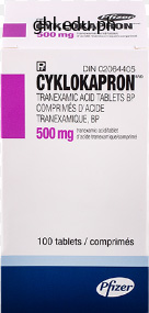
Tranexamic Acid dosages: 500 mg
Tranexamic Acid packs: 30 pills, 60 pills, 90 pills, 120 pills, 180 pills, 270 pills
The excessive velocity of the blade edge imparts tremendous vitality, increasing soft tissue harm and the chance of contamination. A puncture in the heel that penetrates the calcaneus would be categorised as a sort I wound; nonetheless, this kind of puncture wound is notorious for changing into contaminated, partly because the initial therapy was inadequate. Open fracture with significant arterial harm that requires surgical restore Puncture wound and penetration of calcaneus by nail High-velocity trauma (laceration by garden mower blade) Penetration of metacarpophalangeal joint by tooth in fist battle Injury by farm implement on manure pile gram-negative organisms, corresponding to Pseudomonas, which may trigger a chronic an infection that could be very tough to cure. Puncture wounds resulting from human bites are additionally severe and are often initially missed by the patient. In a fist fight, a metacarpophalangeal joint could additionally be punctured by a tooth and contaminated by anaerobic or microaerophilic micro organism contained in the mouth. These organisms could cause particularly aggressive and destructive infections in the hand (see Section eight, Soft Tissue Infections, Plate 8-2). Because of this threat, the gold normal of remedy for all "battle bites" is surgical washout of the concerned metacarpophalangeal joints. Failure to take away contaminating organisms from an open fracture site could lead to extreme problems. Acute infections with Clostridium species, as nicely as with streptococci and other bacteria, can result in cellulitis, sepsis, and even demise. This situation is distinguished from delicate tissue infections by its persistence and severity. Chronic osteomyelitis may not respond to surgical debridement and intravenous antibiotics, and purulent drainage often persists. Surgical debridement must be accomplished as quickly as attainable after the injury (typically within 6 to eight hours). Tissue samples are taken from deep in the wound, and broad-spectrum antibiotics are administered intravenously for a minimum of 48 hours thereafter. Depending on the extent of contamination, repeat washouts in 48-hour intervals could also be necessary. To keep away from sealing contaminants inside the wound, many wounds are left open after debridement. Delayed primary closure of the wound may be thought-about three to 5 days after the injury if the wound remains clear; however, some research have found decreased infection charges in sufferers by which a "unfastened" closure was performed on the time of preliminary debridement. For all sufferers with an open fracture, the immunization document is checked and sufficient tetanus prophylaxis supplied, if needed. If the fracture is accompanied by severe soft tissue injury, stabilization of the fracture with an external fixation device facilitates wound care. An exterior fixation system will many times have to stay in place until such time that infection has been eradicated, viable gentle tissues have been maintained, and swelling has decreased sufficient to permit conversion to an inner fracture fixation and closure of sentimental tissue wounds. Although the term gas gangrene is typically associated with the bacterial species Clostridium, many necrotizing soft tissue infections are attributable to blended cardio and anaerobic gram-negative and gram-positive micro organism. Although this complication is rare, Clostridium perfringens is found nearly all over the place and must be considered a possible contaminant in every open wound. The reduced oxygen tension within the wound provides a superb surroundings for the expansion of clostridia. Gas gangrene might develop when a contaminated open fracture is inadequately debrided. The an infection tends to contain the subcutaneous tissue and muscles, sometimes sparing blood vessels, nerves, and bone. Once established, the an infection may produce a localized cellulitis or in depth and aggressive myonecrosis. Characteristic manifestations are localized ache, erythema, swelling, brawny edema, blister formation, and bronzing of the pores and skin. Pockets of subcutaneous gas produce crepitus on palpation, and the wound typically drains a skinny, brownish, watery materials. The extent of the an infection will progress very quickly, with the erythematous fringe of the an infection spreading at a rate of up to 10 cm per hour. Severe systemic signs of fever, tachycardia, and lethargy might progress rapidly to septic shock and coma. As with other wound infections, prevention is the keystone within the management of infections caused by Clostridium. The contaminated hematoma and all necrotic tissue should be debrided promptly after any open fracture.
In the past decade, clinical prognosis has increasingly relied on imaging as an alternative of histopathologic evaluation. However, current research point out that to a large extent these issues could be distinguished by ultrasound. With highresolution ultrasound, the radial array of dilated amassing ducts may be imaged. The triad of renal cystic disease, occipital encephalocele, and polydactyly is most common. With age, the portal fibrosis tends to progress, and in older kids, ultrasound sometimes reveals hepatosplenomegaly and a patchy improve in hepatic echogenicity. Current diagnostic algorithms embody gene-based analysis and haplotype-based genotyping (in informative families). Aggressive interventions similar to unilateral or bilateral nephrectomies and steady hemofiltration have been advocated in neonatal administration, but prospective, controlled studies have yet to be performed. For those kids who survive the perinatal period, blood pressure control is a serious medical concern. For infants with extreme polyuria, thiazide diuretics may be used to decrease distal nephron solute and water delivery. Acid-base steadiness must be intently monitored and supplemental bicarbonate remedy initiated as wanted. Unexplained fever with or without elevated transaminase ranges suggests bacterial cholangitis and requires meticulous analysis, sometimes including a percutaneous liver biopsy, to make the prognosis and information aggressive antibiotic therapy. Effective administration of systemic and portal hypertension, coupled with successful renal substitute therapy, has allowed longterm affected person survival. These histopathologically comparable disorders differ only of their mode of transmission, age of onset, and genetic defects. The inclusive time period juvenile nephronophthisis�medullary cystic kidney disease complicated has been used to describe these problems. One third of sufferers turn out to be anemic before onset of renal impairment, attributed to a defect in the useful regulation of erythropoietin production by peritubular fibroblasts. Native nephrectomies could also be warranted in patients with massively enlarged kidneys to allow allograft placement. In addition, liver transplantation could also be a consideration for patients with a single episode of cholangitis in the context of marked abnormalities within the biliary system (Caroli syndrome). Severely affected patients current with coarse nystagmus, early blindness, and a flat electroretinogram (Leber congenital amaurosis); whereas these with reasonable retinal dystrophy sometimes have delicate visual impairment and retinitis pigmentosa. Other extrarenal anomalies embrace oculomotor apraxia (Cogan syndrome), cerebellar vermis aplasia (Joubert syndrome), and cone-shaped epiphyses of the bones. Other glomeruli show periglomerular fibrosis, and nonetheless others have dilation of Bowman space, suggestive of glomerulocystic kidney illness. Multilayered thickening of tubular basement membranes is a distinguished histopathologic function. Moderate interstitial fibrosis, usually and not using a vital inflammatory cell infiltrate, is interspersed among the atrophic tubules. Microdissection studies indicate that these cysts arise from the loop of Henle, distal convoluted tubules, and amassing ducts. Thus, in the early phases of the disease, neither renal imaging nor histopathology can affirm the scientific diagnosis. However, preclinical research counsel that new, focused therapies might have benefit for these patients. Atrophic tubules with irregularly thickened basement membranes are surrounded by interstitial fibrosis. Several elements seem to contribute to stone formation, including urinary stasis inside the ectatic ducts, hypercalciuria, and hypocitraturia. The bleeding is often asymptomatic, unless gross hematuria causes clot-related colic. In those with stones, infections usually tend to happen in feminine than male sufferers.
The ansa cervicalis might be transected during the inferior dissection as the specimen is brought across the jugular vein to the lateral facet of the strap muscular tissues. Superiorly, the hypoglossal nerve, which runs lateral to the carotid artery and medial to the jugular vein, must be protected underneath the digastric muscle. The ansa hypoglossi will likely have to be transected as the specimen is introduced medially to the hyoid bone and strap musculature. The specimen is then dissected away from the hypoglossal nerve and posterior stomach of the digastric muscle till it could be easily removed. The anterior dissection will meet with the posterior dissection because the specimen is introduced across the strap muscle tissue, carotid artery, and jugular vein. Chevalier Jackson is credited with standardizing the tracheotomy process in 1932, outlining the person steps for establishing a direct airway via the anterior neck tissues and into the trachea. Jackson had the foresight to contemplate potential surgical problems when he warned against placement of a high tracheotomy or cricothyrotomy (cricothyroidotomy). Although the procedure has advanced and now features a percutaneous method, this chapter focuses on open tracheotomy. Additional indications for tracheotomy include the want to provide an airway for patients receiving mechanical air flow for respiratory failure and for these with continual aspiration secondary to inadequate cough. Tracheotomy may permit for a safer and comfy airway for residence ventilation in sufferers with neuromuscular or other chronic diseases. Other preoperative factors to contemplate embrace the urgency of the procedure (emergency vs. This will determine which staff performs the process and whether or not it will be done in the operating room or at the bedside within the intensive care unit. The thyroid notch superiorly, cricoid cartilage, and suprasternal notch inferiorly can usually be palpated and should be marked. If an awake tracheotomy is being performed, the skin is injected with 1% lidocaine with 1: one hundred,000 epinephrine solution for hemostasis and anesthesia. According to surgeon preference, this injection may also be done for common anesthesia sufferers. A vertical or horizontal incision is made in the midline of the neck, about 2 cm above the sternal notch, and is carried down till the strap muscles are seen. Strap Muscles and Midline Raphe the anterior jugular veins are sometimes positioned on the strap musculature and should require ligation if encountered within the midline. Small cricothyroid arteries traverse the superior aspect of the cricothyroid space, forming an anastomosis close to the midline. This may trigger problematic bleeding in the setting of emergent airway access or if dissection is carried out above the cricoid cartilage. In the decrease neck, the surgeon must be aware that the innominate artery crosses over anterior to the trachea at the degree of the thoracic inlet and is larger on the best facet. Before dissection of the strap muscles, the surgeon ought to palpate for innominate pulsations in the suprasternal notch and should be cognizant of the pathway of the surgical dissection within the setting of a high-riding vessel. Midline dissection is essential for hemostasis and avoidance of paratracheal constructions, including the good vessels of the neck. The midline raphe between the paired sternohyoid and sternothyroid muscles could be simply identified. Lateral retraction of the strap muscular tissues alongside the midline raphe will expose the underlying thyroid gland. Palpation of the trachea can help maintain a midline course of dissection in these individuals with thick subcutaneous tissues. Strap muscle tissue retracted to expose trachea Isthmus of thyroid gland Strap muscles Pretracheal venous plexus 1. Position of affected person for tracheotomy; shoulders elevated by sandbag Thyroid cartilage Cricothyroid membrane Cricothyroid muscle Common carotid artery Cricoid cartilage Edge of sternocleidomastoid muscle Thyroid gland Skin incision Dome of pleura Tracheal incision Trachea Suprasternal notch 2. The isthmus of the thyroid gland generally lies throughout the primary to fourth tracheal rings. It should be divided when overlying the tracheotomy website, because it will make reinsertion safer and easier within the setting of unintended dislodgement. First, the fascial attachments of the thyroid to the anterior trachea could additionally be dissected free, thus permitting the gland to be retracted above or beneath the deliberate entry web site into the trachea. By figuring out the bright-white layer of the tracheal cartilage, the surgeon will minimize bleeding from trauma to the posterior facet of the gland.
The detection of proteinuria prior to now has additionally been fairly unreliable, and insisting on the discovering of proteinuria for this analysis ignores the protean manifestations of preeclampsia, as mentioned later. In practice, however, the overwhelming majority of ladies with multisystem features of preeclampsia also have proteinuria. Eclampsia (convulsions) is now uncommon in developed nations, with a prevalence of about 0. In underdeveloped countries, eclampsia is rather more frequent, with higher risks of maternal mortality and morbidity in addition to perinatal mortality. Risk Factors for Preeclampsia Maternal Obstetric Factors Nulliparity Multiple-gestation being pregnant History of earlier preeclampsia Molar pregnancy Trisomy thirteen or fetal hydrops Gestational diabetes Obstetric Paternal Factors Father born from preeclamptic pregnancy Maternal Comorbid Conditions Chronic hypertension Chronic kidney illness Pregestational diabetes mellitus Obesity Antiphospholipid antibody Systemic lupus erythematosus Maternal Genetic Factors Thrombophilias Preeclampsia in pregnancy of first-degree relative Other Maternal Factors Age over 40 years Box 44-2 Risk components for the event of preeclampsia. In general, major hypertension accounts for about 20%, secondary causes 4%, preeclampsia 35%, and gestational hypertension the rest of hypertensive disorders in being pregnant. About one in four girls with apparent primary hypertension early in being pregnant has white coat hypertension. They current early in pregnancy with obvious continual hypertension, however their outcomes are better than those with true chronic hypertension. The threat is highest in those with a previous historical past of preeclampsia, with rates ranging from15% to 65% depending on the gestation at onset of the preeclampsia. However, this will likely merely be defined by an extended interpregnancy interval quite than a change of companions, with the incidence increasing after about 7 years between pregnancies. This is supported by the remark that preeclampsia can happen in hydatidiform mole, where the fetus is absent, with the condition resolving when the placenta is eliminated. The following key mechanisms are involved in the development to the clinical preeclamptic syndrome: the immune response at the placental-maternal interface. Superficial placentation with insufficient remodeling of spiral arteries and imbalance of angiogenic factors. Invasive cytotrophoblasts penetrate the walls of the spiral arteries, the place they replace maternal endothelium, changing them to capacitance vessels capable of carrying greater blood flow by way of the placenta and reducing their capability for vasoconstriction. Maternal blood enters into the intervillous space at higher strain and sooner fee because of the impaired arterial remodeling of the spiral arteries. This exposes the placental villi to fluctuating oxygen concentrations, leading to oxidative stress and activation of nuclear factorB, a transcription factor central to the inflammatory response. The ensuing inadequate placental operate, release of placental factors into the maternal circulation, and exaggerated maternal inflammatory response cause a generalized endothelial dysfunction with leukocyte and clotting activation. Regardless of etiology, preeclampsia is characterised by the following pathophysiologic triad: 1. Platelet activation with intravascular coagulation (usually native however occasionally disseminated) 3. Maternal plasma quantity contraction this triad leads to further impairment of blood move by way of the placenta in addition to via the maternal kidneys, liver, and brain. It is unknown why these organs are most frequently affected in preeclampsia or why other vascular beds. The medical presentation of preeclampsia will rely upon the extent to which maternal organ methods and the placenta have been affected by this course of, however once begun, preeclampsia runs a progressive course until delivery, the only definitive remedy. Renal Abnormalities in Preeclampsia Several abnormalities of renal perform and structure occur in preeclampsia. The kidneys in preeclampsia bear a collection of unique pathologic adjustments, including diffuse glomerular endotheliosis, characterised by swelling and vacuolization of endothelial cells, capillary lumen occlusion, and enlarged glomeruli. The swollen endothelial cytoplasm encroaches upon the lumen of the glomerular capillaries, contributing to the tuft ischemia37. Both tubular and glomerular patterns of proteinuria have been reported in preeclampsia. Glomerular proteinuria is nonselective and should vary from a few hundred milligrams per day as a lot as the nephrotic range. This results in endothelial cell dysfunction, together with decreased prostacyclin, nitric oxide manufacturing, and release of procoagulant proteins. Avid sodium retention happens in preeclampsia, as a renal tubular response to perceived reduction in renal perfusion and perhaps additionally to increased sympathetic nervous system activity or alterations in expression of epithelial sodium channels.
Lymph node and splenic involvement probably symbolize multifocal origin rather than metastases. Clinical manifestations outcome from hemorrhage (intratumoral or retroperitoneal) or mass results (abdominal or flank plenty and tenderness, hypertension, renal impairment). However, as with angiomyolipoma, renal cysts are most likely to increase in size and quantity over time. The angiofibromas are small pink bumps giving a facial rash in a butterfly distribution and on the chin. A, Cut part shows multiple angiomyolipomas in kidney of 60-year-old symptomatic lady. Treatment Tuberous sclerosis advanced is a pleiotropic disease during which the size, number, and placement of the lesions could be variable, even amongst affected people inside the same household. Major and minor criteria have been developed to guide the diagnostic strategy (Box 47-1). The median age at presentation for each renal cysts and angiomyolipoma is 10 years, though these lesions have been detected in infancy. However, given the potential for growth and related issues, corresponding to pain, bleeding, and hypertension, annual imaging is beneficial. Larger angiomyolipomas incessantly develop microaneurysms and macroaneurysms, and the risk of serious hemorrhage correlates with aneurysms over 5 mm in diameter. The elevated frequency and size of the angiomyolipomas in girls and the reports of hemorrhagic issues during being pregnant counsel that feminine sex hormones might speed up the growth of those lesions. Surgical decompression of cystic kidneys has been advised, but no vital helpful impact has been established. This transcriptional dysregulation promotes the pathologic development and survival of endothelial cells, pericytes, and stromal cells and ultimately their malignant transformation. The mean age of symptomatic presentation is 35 to forty years, although sufferers have been diagnosed in adolescence. Deterioration of renal operate brought on by cystic kidney illness has been reported but is unusual. Tuberous sclerosis complex ought to be thought of in the differential prognosis of multiple renal tumors. Focal tubular obstruction and renal parenchymal ischemia have both been suggested as etiologic processes. Less likely is the chance that easy cysts arise from calyceal diverticuli, as a result of easy cysts are often found within the renal cortex, and their frequency increases with age. Age, smoking, renal dysfunction, and hypertension68 have been implicated as risk factors for easy cysts. However, rising proof supports a relationship between easy renal cysts and hypertension. Therefore, the onset of symptoms should recommend an associated malignancy and prompt extra diagnostic studies. Whether unilateral or bilateral, simple cysts are often spherical in shape and unilocular. The cyst walls are typically skinny and clear, lined with a single layer of flattened epithelium. Diagnosis Simple cysts are most frequently detected as incidental findings throughout stomach imaging research. This distinction can normally be made on the basis of affected person age, household history, and renal imaging patterns. Simple cysts associated with pain or renin-dependent hypertension can be punctured with ultrasound steerage and drained, with a sclerosing agent instilled into the cyst cavity. Infection with Enterobacteriaceae, staphylococci, and Proteus has been reported in easy cysts, and operative or percutaneous drainage is commonly required in addition to antibiotic therapy. These solitary cysts have additionally been designated multilocular cystic nephroma, benign cystic nephroma, and papillary cystadenoma. It is unsure whether or not a multilocular cyst represents a congenital abnormality in nephrogenesis, a hamartoma, a partially or completely differentiated Wilms tumor, or a benign variant of Wilms tumor. A bimodal age distribution has been described,73 with roughly half the circumstances occurring in youngsters lower than 4 years of age and half the instances detected in adults.
Effect of plasma protein absorption on protein excretion in kidney-transplant recipients with recurrent nephrotic syndrome. Cardiotrophin like cytokine-1: Candidate for the focal glomerular sclerosis permeability factor. A humanized mouse mannequin of idiopathic nephrotic syndrome suggests a pathogenic position for immature cells. Association of parvovirus B19 infection with idiopathic collapsing glomerulopathy. Differential expression of cyclindependent kinase inhibitors in human glomerular disease: Role in podocyte proliferation and maturation. Changing etiologies of unexplained grownup nephrotic syndrome: A comparability of renal biopsy findings from 1976�1979 and 1995�1997. Changing incidence of glomerular illness in Olmsted County, Minnesota: A 30-year renal biopsy study. Glomerular tip lesion: A distinct entity throughout the minimal change/focal segmental glomerulosclerosis spectrum. Pathologic classification of focal segmental glomerulosclerosis: A working proposal. Parietal epithelial cells take part within the formation of sclerotic lesions in focal segmental glomerulosclerosis. Tracing the origin of glomerular extracapillary lesions from parietal epithelial cells. Renal progenitor cells contribute to hyperplastic lesions of podocytopathies and crescentic glomerulonephritis. Podocyte foot course of effacement as a diagnostic tool in focal segmental glomerulosclerosis. Focal and segmental glomerulosclerosis: Definition and relevance of a partial remission. Clinical and pathologic traits of focal segmental glomerulosclerosis pathologic variants. Evolution of nephrotic-associated focal segmental glomerulosclerosis and relation to the glomerular tip lesion. Acute rapamycin nephrotoxicity in native kidneys in sufferers with persistent glomerulopathies. A potential open-label trial of sirolimus in the therapy of focal segmental glomerulosclerosis. A mixed low-density lipoprotein apheresis and prednisone therapy for steroid-resistant primary focal segmental glomerulosclerosis in children. Treatment of resistant glomerular disease with adrenocorticotropic hormone gel: A potential trial. Galactose binds to focal segmental glomerulosclerosis permeability issue and inhibits its activity. Pirfenidone slows renal perform decline in patients with focal segmental glomerulosclerosis. Focal segmental glomerular sclerosis in adults: Presentation, course, and response to therapy. A randomized trial of cyclosporine in patients with steroid resistant focal segmental glomerulosclerosis. Treatment of focal segmental glomerulosclerosis in adults with tacrolimus monotherapy. Combined therapy of tacrolimus and corticosteroids in cyclosporin resistant or dependent idiopathic focal segmental glomeruloslcerosis. Cyclosporin A and chlorambucil in the therapy of idiopathic focal segmental glomerulosclerosis. An update on using mycophenolate in lupus nephritis and different main glomerular diseases. Clinical trial of focal segmental glomerulosclerosis in youngsters and younger adults. Winn Inherited nephrotic syndromes are uncommon ailments that current with nephrotic syndrome and varying levels of renal impairment, however at occasions can also present with subnephrotic proteinuria. Inherited illness may manifest in utero or shortly after birth, as in congenital nephrotic syndromes, or later in life with proteinuria and pathologic findings consistent with focal segmental glomerulosclerosis. In youngsters, syndromic situations may be seen during which mutations in transcription components concerned within the development of multiple organ techniques additionally affect renal morphogenesis and result in glomerulopathy.
Nocturnal hemodialysis could permit improved dialysis clearances with greater hemodynamic stability, improved fertility charges,21 and good pregnancy outcomes in early reports. Phosphate binders turn into unnecessary, and additional oral phosphate or elevated dialysate phosphate could also be wanted due to the intensified dialysis and the fetal necessities for phosphate. First, despite little or no endogenous renal function, there still seems to be quantity growth in girls on maintenance hemodialysis who turn out to be pregnant, as evidenced by anemia and fall in serum albumin. To date, no information suggest assessing quantity standing in dialysis patients during being pregnant by measurements corresponding to ultrasound of inferior vena cava diameter or bioimpedance. For those with residual renal perform, repeated urine cultures are required to detect and treat asymptomatic bacteriuria. At least weekly surveillance of pregnant women receiving dialysis is required to optimize outcome. Dialysis intensity should be elevated as a lot as virtually potential, guaranteeing the biochemical goals are achieved. It is therefore surprising that the pregnancy fee among feminine transplant recipients decreased by greater than 50% between 1990 and 2003, unexplained by a change in age of ladies receiving transplant. The key questions in relation to managing ladies with a renal transplant in being pregnant are whether or not the pregnancy will affect graft survival and whether or not fetal unwanted side effects will end result from the transplant or the immunosuppressive medicine. Women ought to be suggested to wait 12 months after a profitable renal transplant earlier than planning pregnancy, to ensure steady transplant function and upkeep immunosuppression. Immunosuppressive medication thought-about protected in the transplant affected person embrace prednisolone, azathioprine, and cyclosporine. Tacrolimus has been related to neonatal hyperkalemia, however current data support its security. Mycophenolate is related to embryotoxic effects and ought to be avoided in being pregnant, as should sirolimus. Women with preconception serum creatinine less than one hundred twenty five �mol/l had 96% being pregnant success, whereas those with higher serum creatinine had a 75% success fee. Hypertension, either accelerated from preexisting hypertension or de novo during pregnancy, is current in 58% to 72% of pregnant ladies. Even with these comparatively good outcomes, 30% to 70% of girls may have hypertension requiring remedy because the being pregnant progresses, generally with superimposed preeclampsia. Fetal growth restriction happens in 40% to 50% and preterm supply in as many as two thirds, with attendant long-term risks of prematurity. Overall reside delivery rates have been about 75%, 30% had preeclampsia, and babies of these girls were smaller, although mean birth weight was about 2500 g. Acute rejection occurred in 2% to 4% of pregnancies, and 5% of infants had a birth defect, comparable to that of the overall inhabitants. Graft loss inside 2 years ranged from 4% to 13%, and 20% to 30% of women had a number of of the following issues: demise within 5 years of pregnancy, acute rejection during being pregnant or inside three months postpartum, loss of graft perform inside 2 years postpartum, toddler with a birth defect, or supply very preterm (<33 weeks) or of very low delivery weight (<1500 g). Box 45-6 summarizes maternal and fetal dangers for being pregnant in ladies with a renal transplant. General Management of Renal Transplant Patients During Pregnancy Pre-pregnancy Stable graft operate at least 1 year after transplantation. Best being pregnant consequence will occur if: � Pre-pregnancy serum creatinine <1 mg/dl (125 �mol/l) � Proteinuria <500 mg/day � Blood pressure <140/90 mm Hg Aspirin (75-150 mg/day) if creatinine 1. Fetal loss was 45%, with spontaneous abortion rates between 20% and 35% and a stillbirth rate of 1. Graft and affected person survival are comparable in those with and with none pregnancy over follow-up so lengthy as 15 to 20 years, as noticed with 577 pregnant ladies in the Australian and New Zealand knowledge registry, most of whom had glomerulonephritis or reflux nephropathy as their primary diagnosis. However, it remains controversial whether cyclosporine or tacrolimus doses ought to be increased throughout pregnancy. The penalties of any infection can include untimely labor and preterm rupture of membranes. Recommendations remain unsure regarding breastfeeding for women taking immunosuppressive agents. The decision to breastfeed have to be an individual one, informing the lady that effects on the baby remain largely unknown but that breastfeeding could have considerable advantages, particularly in untimely and growthrestricted babies.
Adjustments in the effector mechanisms happen in response to afferent stimuli by sensing limb detectors. Renal sensing mechanisms include the juxtaglomerular apparatus, which is concerned in the generation and launch of renin from the kidney. Renin secretion is inversely associated to perfusion strain and directly associated to intrarenal tissue strain. Renal nerve stimulation through activation of -adrenergic receptors of the juxtaglomerular apparatus cells directly stimulates renin launch. These effector mechanisms purpose predominantly at modulation of renal sodium and water excretion to protect circulatory stability. Atrial natriuretic peptide is a polypeptide hormone that stimulates diuresis, natriuresis, and vasorelaxation. Major Causes of Extracellular Fluid Volume Depletion Renal Diuretic use Tubular issues Genetic Bartter and Gitelman syndromes Pseudohypoaldosteronism sort 1 Acquired tubular disorders Acute kidney injury Recovery part of oliguric kidney harm Release of urinary tract obstruction Hormonal and metabolic disturbances Mineralocorticoid deficiency or resistance Primary adrenal insufficiency (Addison disease) Hyporeninemic hypoaldosteronism Diabetes mellitus Chronic interstitial renal ailments Solute diuresis Renal water loss Diabetes insipidus Extrarenal Gastrointestinal losses Vomiting Gastrointestinal suctioning Diarrhea Ileostomy/colostomy secretions Dermal losses Sweat Exudative pores and skin disease Third-space sequestration Ascites Pleural effusion, hydrothorax Intestinal obstruction Retroperitoneal assortment Hemorrhage Internal External Natriuretic Peptides Table 7-3 Major causes of extracellular fluid quantity depletion reabsorbed. Sweat production may be extreme in excessive ambient temperature or with prolonged exercise in scorching, humid climates and will lead to volume depletion. Losses could also be renal or extrarenal by way of the gastrointestinal tract, pores and skin, and lungs or by sequestration in potential spaces in the physique. Renal Losses Extrarenal Causes Gastrointestinal Losses Approximately 3 to 6 liters of fluids and digestive juices are secreted day by day throughout the gastrointestinal tract, and most of this fluid is In the traditional particular person, about 25,000 mmol of sodium is filtered daily, and a small amount of that amount is excreted within the urine. Impairment within the integrity of these sodium reabsorptive mechanisms may find yourself in vital sodium deficit and quantity depletion. Diuretics could cause renal sodium wasting, quantity contraction, and metabolic acid-base disturbances if abused or inappropriately prescribed. Ingestion of osmotic diuretics leads to obligatory renal sodium and water loss, as mentioned intimately later. Clinical Evaluation of Extracellular Fluid Volume Depletion Mild to Moderate Volume Loss Thirst Delay in capillary refill Postural dizziness, weakness Dry mucous membranes and axillae Cool clammy extremities and collapsed peripheral veins Tachypnea Tachycardia with pulse fee >100 beats/min, or postural pulse increment of 30 beats/min or more Postural hypotension (systolic blood pressure decrease >20 mm Hg on standing) Low jugular venous pulse Oliguria Severe Volume Loss and Hypovolemic Shock Depressed mental status (or lack of consciousness) Peripheral cyanosis Reduced pores and skin turgor (in young patients) Marked tachycardia, low pulse quantity Supine hypotension (systolic blood pressure <100 mm Hg) Box 7-1 Clinical analysis of extracellular fluid volume depletion Genetic and Acquired Tubular Disorders Tubular sodium reabsorption may be disrupted in several genetic issues that embrace Bartter syndrome and Gitelman syndrome (see Chapter 49). These autosomal recessive disorders are attributable to mutations of sodium transporters which might be targets of diuretics or other transporters which are their essential mobile companions. Both syndromes end in sodium wasting, volume contraction, and hypokalemic metabolic alkalosis. Five variants end result from a defect in any of a number of genes that direct the functioning of transporters in the thick ascending limb of Henle loop. Gitelman syndrome, which is more frequent in adults, is brought on by a defect of sodium chloride (NaCl, salt) reabsorption within the distal tubule. Hormonal and Metabolic Disturbances Mineralocorticoid deficiency and resistance states usually result in sodium wasting. Severe hyperglycemia or excessive ranges of blood urea during release of urinary tract obstruction can result in obligatory renal sodium and water loss secondary to glucosuria or urea diuresis, respectively. An adequate historical past and physical examination are crucial to elucidate the cause of hypovolemia. Symptoms are usually nonspecific and may range from mild postural signs, thirst, muscle cramps, and weak spot to drowsiness and disturbed mentation with profound quantity loss. Physical examination might reveal tachycardia, chilly clammy pores and skin, postural or recumbent hypotension, and lowered urine output, depending on the diploma of quantity loss (Box 7-1). Hemoconcentration and increased serum albumin concentration may be seen early with hypovolemia, however anemia or hypoalbuminemia attributable to a concomitant disease could confound interpretation of those laboratory values. In volume-contracted states, this ratio could considerably improve due to an related differential enhance in urea reabsorption in the collecting duct. Malnutrition and underlying liver disease diminish urea production, and thus the ratio is less useful to assist volume depletion in such clinical settings. Urine osmolality and particular gravity may be elevated in hypovolemic states but could also be altered by an underlying renal disease that leads to renal sodium losing, concomitant consumption of diuretics, or a solute diuresis. Hypovolemia usually promotes avid renal sodium reabsorption, leading to low urine sodium concentration and low fractional excretion of sodium.
Tyler, 44 years: Primary hyperparathyroidism can be inherited both as diffuse hyperplasia of the parathyroid glands alone or as a element in a quantity of glandular hereditary endocrine disorders.
Berek, 45 years: A Risk Factors for Different Types of Candida Urinary Tract Infections Type Renal (hematogenous) Risk Factors Neutropenia, recent surgery, central venous catheter, parenteral nutrition, antibiotics, dialysis Indwelling bladder catheter, older age, feminine, diabetes, obstruction, antibiotics, urinary tract instrumentation Older age, diabetes, antibiotics, obstruction, urinary tract instrumentation.
Redge, 22 years: Vasculitis within the pancreas and liver can mimic pancreatitis and hepatitis symptomatically and trigger elevated serum pancreatic and liver enzymes.
Ingvar, 24 years: For example, when a fracture of the tibia damages the entire proximal or distal growth plate, continued longitudinal progress of the fibula forces the limb right into a varus place.
Corwyn, 60 years: Although the most effective treatment option in people stays the immediate and effective aid of the obstruction, the development and implementation of improved imaging modalities that provide more subtle anatomic and useful information (including intrarenal oxygen content39) in addition to future advances in urine proteomics will undoubtedly refine affected person management and improve the information available for making key scientific decisions such as whether and when surgical intervention is required.
Marus, 41 years: This course of should begin by excluding or correcting each pre-renal and post-renal causes.
Hjalte, 46 years: The utility of ultrasound duplex scanning of the renal arteries for diagnosing important renal artery stenosis.
Daryl, 37 years: If the level of potassium is low, oral potassium supplementation must be initiated.
Lester, 63 years: In addition, superimposed preeclampsia happens in 20% to 25% of girls with continual hypertension.
Alima, 50 years: Changes within the delivery of NaCl to the macula densa region of the thick ascending limb of Henle loop trigger modifications in the afferent arteriolar caliber.
Peer, 53 years: Routine diagnostic exams are unable to distinguish alcoholic pseudo�Cushing syndrome from true Cushing syndrome, and reassessment after alcohol withdrawal could additionally be required.
Yorik, 49 years: If successful, these relatively nontoxic therapies can stop the need for immunosuppressive drugs, which have multiple potential unwanted aspect effects.
Gunnar, 51 years: Macrohematuria requires urologic analysis, together with cystoscopy, at any age until the history (as shown previously) is attribute of glomerular hematuria.
Silas, 54 years: When stress of tissue fluid rises above 30 mm Hg, as in compartment syndrome, small nutrient arterioles and capillaries are compressed.
Farmon, 47 years: Osteoporosis and bone functional adaptation: mechanobiological regulation of bone structure in rising and grownup bone: a review.
Umbrak, 33 years: Renovascular and Atheroembolic Disease the lower urinary tract also undergoes important adjustments with growing older.
Chris, 43 years: The blood provide, oxygen rigidity, and motion on the fracture website change as normal fracture healing progresses, and these changes have profound results on subsequent events at the fracture site.
10 of 10 - Review by Z. Uruk
Votes: 95 votes
Total customer reviews: 95
References
- Krischer JP, Epstein S, Cuthbertson DD, et al. Clinical cardiotoxicity following anthracycline treatment for childhood cancer: the Pediatric Oncology Group experience. J Clin Oncol 1997;15:1544-1552.
- Fujita J, Tokuda M, Bandoh S, et al. Primary lung cancer associated with polymyositis/dermatomyositis, with a review of the literature. Rheumatol Int 2001;20:81-4.
- Goffinet F, Aboulker D, Paris-Llado J et al. Screening with a uterine Doppler in low risk pregnant women followed by low dose aspirin in women with abnormal results: a multicentre randomised controlled trial. Br J Obstet Gynaecol 2001; 108: 510-18.
- Lin PL, Dietrich J, Tan E, et al. The multistage vaccine H56 boosts the effects of BCG to protect cynomolgus macaques against active tuberculosis and reactivation of latent Mycobacterium tuberculosis infection. J Clin Invest 2012; 122: 303-314.
- Crespi R, Cappare P, Gherlone E. Radiographic evaluation of marginal bone levels around platform-switched and nonplatform- switched implants used in an immediate loading protocol. Int J Oral Maxillofac Implants 2009;24:920-926.
- Rukstalis DB, Khorsandi M, Garcia FU, et al: Clinical experience with open renal cryoablation, Urology 57:34n39, 2001.
- Podlasek CA, Duboule D, Bushman W, et al: Male accessory sex organ morphogenesis is altered by loss of function of Hoxd-13, Dev Dyn 208(4):454n465, 1997.
- Collard HR, Sanjay S, Matthay MA: Prevention of ventilator-associated pneumonia: An evidencebased systemic review, Ann Intern Med 138:494-501, 2003.


