
Bernardino D. Madsen, MT (ASCP)
- Instructor
- Medical Laboratory Technology Program
- Casper College
- School of Health Science
- Casper, Wyoming
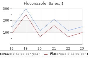
Fluconazole dosages: 400 mg, 200 mg, 150 mg, 100 mg, 50 mg
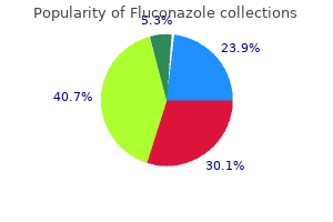
Fluconazole packs: 30 pills, 60 pills, 90 pills, 120 pills, 180 pills, 270 pills, 360 pills

Fluconazole 50 mg on line
Transient visual loss has been reported after posterior capsular tear in instances of intracameral lidocaine use. When topical anesthesia is getting used it could be very important clarify to the patient every step of the process and the anticipated feeling. Modes of Anesthesia Topical Advantages Anesthesia No perforation Rapid visible recovery No injection ache No akinesia Some discomfort (pain) Intracameral Iris manipulation Less affected person discomfort Subconjunctival Anesthesia Less Peribulbar Anesthesia Akinesia Avoid some retrobulbar problems Complications: globe penetration, muscle damage Retrobulbar Anesthesia Akinesia General Sparing affected person cooperation Disadvantages No akinesia No akinesia Major issues: orbital hemorrhage, Optic nerve/ muscle damage Anesthesia dangers. The needles are directed parallel to the orbital partitions in the location comparable to the superior and inferior lateral third of the orbital rim. Eyelid akinesia may be achieved by 1�2 mL anesthetic injection from the lateral side, alongside and adjoining to the rim and parallel to the surface. The anesthesia and akinesia are normally quick, but attainable complications embrace inadvertent penetration of the globe (especially in myopic eyes) or the optic nerve, injection to the optic nerve sheath, muscle damage, retrobulbar hemorrhage, oculocardiac reflex, and potential systemic toxicity and allergic reactions. In addition to these modes, common anesthesia is indicated for uncooperative patients. The needle is launched instantly superior to the lateral third of the inferior orbital rim. The needle is passed parallel to the orbital flooring and angled superomedially instantly past the globe equator. When viscoelastic agent is injected into the anterior chamber, the maintainer and irrigating fluid must be closed. The primary surgical wound ought to be customary earlier than hydrodissection and hydrodelineation are performed and they should be performed through this incision. Paracenteses are used to inject viscoelastic agents, performing capsulorrhexis, or insertion of chopper and spatula. All cannulas which might be inserted into the eye should be full of a small quantity (0. If intraocular bleeding happens during surgery, elevation on the infusion bottle connected to an anterior chamber maintainer must be sufficient. Air bubbles in the anterior chamber could also be aspirated with the phaco tip in position 2 (aspiration/ irrigation) and never in place 3 (phacoemulsification) or with the aspiration/irrigation tip. The advantages of anterior chamber maintainer are that it preserves the depth of the anterior chamber in case of inadvertent sudden surge and it additionally allows infusion of agents into the attention. The paracentesis is directed barely posterior and obliquely to be able to keep away from move toward the endothelium or the capsulorrhexis margins. The maintainer could also be inserted firstly of the surgical procedure with its opening confronted posteriorly to keep away from water jet injury to the endothelium. Additional one or two paracenteses are often carried out between 2 and 3 clock hours from and on either side of the primary incision to enable injection of viscoelastic agent or the introduction of a second instrument in bimanual cataract extraction. Such incisions promote extra fast healing, reduce astigmatism, scale back potential infections, and supply speedy visible rehabilitation. A temporal incision is usually used if against-the-rule astigmatism is present before surgery. Access to the anterior chamber is easier via a temporal incision due to the shallow lateral orbital wall. Three patterns of external incisions are generally employed; the standard curvilinear, the straight, and the frown incisions. The opposite-shaped frown incision theoretically presents the best help and induces the least amount of astigmatism. Induced astigmatism is estimated to be proportional to the cubic length of such incisions. The internal opening of the incision into the anterior chamber additionally influences the amount of induced astigmatism. The scleral (or sclerocorneal) tunnel connecting the exterior and inside openings may have different configurations. If the tunnel is directed toward the anterior chamber, it creates a biplanar incision. A vertical 300- to 500-mm incision is made and prolonged as a scleral tunnel parallel to the floor. The anterior chamber is penetrated by directing the blade posteriorly, creating the third plane.
Order 200 mg fluconazole free shipping
The racial steadiness of these sufferers in all probability correlates with the racial composition of the catchment inhabitants in this examine. After an exhaustive evaluation of the literature, they concluded that the number of reported familial circumstances was too small to show a relation beyond coincidence. The most typical signs on the time of presentation are decreased imaginative and prescient or glare because of cataract formation. Other patients could present with gentle ocular discomfort or ciliary spasm-type ache, although associated conjunctival injection and photophobia are comparatively uncommon. Occasional sufferers seek medical consideration because they detect heterochromia or a change in iris colour. Uncommon instances of symptoms attributable to elevated intraocular pressure, spontaneous hyphema, and strabismus from juvenile cataract have been reported. In a big proportion of cases, however, a number of of the three basic signs could additionally be missing. Fuchs emphasized the presence of heterochromia, cataract, keratic precipitates, and different medical features. In some patients, the heterochromia was marked, whereas in others, it was extraordinarily refined. In other sufferers, a change in iris colour was detected later in life when visual impairment was current. Fuchs additionally observed that the pupil was sometimes enlarged and poorly reactive to light and lodging. Keratic precipitates had been present in all instances examined with magnification, present in no less than 30 (79%) of 38 sufferers. In cases in which the vitreous could probably be observed, vitreous opacities had been incessantly present. He confused the distinct lack of overt indicators of inflammation, such as ache, ciliary injection, photophobia, and miosis. In most instances, loss of pigment from the anterior border layer and stroma leads to hypochromia of the affected eye. In blue-eyed sufferers, the affected eye often seems more intensely blue or lighter in shade than the other eye. This occurs on account of the lack of the orange-brown pigment of the anterior border layer, which is normally extra dense around the collarette. Subtle heterochromia is best detected utilizing pure daylight or bright overhead lighting. This is best achieved with the unaided eye; slight differences in iris shade are tough to detect by slit lamp. Perhaps the most delicate methodology for detecting heterochromia is to evaluate anterior section images taken under standardized circumstances. Tips Use pure mild or bright overhead lighting to detect refined heterochromia. Slight differences in iris color are tough to detect by slit lamp examination. A small variety of sufferers are able to identify the age at which heterochromia was acquired. The profound hypochromia in the left eye is because of loss of anterior border layer pigment. The apparent hyperchromia of the proper (affected) iris is as a end result of of anterior border layer and stromal atrophy revealing the underlying iris pigment epithelium. One of essentially the most striking features is the depigmentation of the anterior border layer. This structure is a condensed layer of stromal cells at the anterior surface of the iris instantly beneath the endothelial masking. In darkish brown eyes, this layer is richly pigmented and imparts the traditional velvety appearance to the floor of the brown iris. In blue irides, this layer is relatively translucent and divulges the underlying stromal structure, together with normal iris vessels. This condensation is extra dense across the collarette and is answerable for the speckled orange�brown pigment noticed in some blue eyes. Transillumination defects are usually scattered but could have a predilection for the pupillary region. Infrequently, iris epithelial pigment deposits may be detected on the anterior lens capsule, probably representing a web site of previous synechiae formation.
Fluconazole 400 mg otc
Another example is sickle cell anemia by which heterozygotes are protected against the extreme pathogenesis of malaria. Genetic proof signifies that every one vertebrate opsin genes developed from a standard ancestor via a means of gene divergence and duplication. In New World primates, trichromatic shade vision was acquired by way of evolution of allelic variety in the X-chromosome opsin gene, which produced selection in spectral sensitivity of the encoded photopigments. In Old World primates, trichromatic color imaginative and prescient arose via a gene duplication that placed two opsin genes together in tandem on the X-chromosome. Calling them short-, middle-, and long-wavelength sensitive, abbreviated S, M, and L cones minimizes confusion that may come up from giving them colour names. Photopigment molecules within each cone are answerable for the spectral properties of the cones. Each photopigment molecule is composed of two parts; a protein termed the opsin, and an 11-cis-retinal chromophore. The noncontributing cone class is indicated by the prefixes: Protan- for absence of L cone contribution to vision. Further categorization of colour vision defects is dependent upon whether or not the remaining colour imaginative and prescient relies on two (dichromacy) versus three (anomalous trichromacy) spectrally distinct types of cones. The suffix -anomaly denotes anomalous trichromacy by which two of the cone courses are more comparable in spectral sensitivity than the corresponding regular cones: Deuteranopia. The number of regular and defective colour imaginative and prescient phenotypes in humans is produced by unequal homologous recombination during meiotic cell division in females. The shade of the arrowhead indicates whether the gene encodes an L pigment (red) or M pigment (green). Only the two 5, genes (left-most) in the three gene array are expressed and each of those encode L-class pigments. The array produces dichromatic shade imaginative and prescient if the encoded L-class pigments have equivalent spectral properties, or it produces anomalous trichromacy if the pigments differ in spectral properties. The different recombinant array contains a single gene, which encodes an L opsin and thus produces dichromatic color imaginative and prescient. As might be mentioned below, the diversity introduced by intermixing L and M opsin gene sequences underlies the number of phenotypes related to anomalous trichromacies. Only amongst humans is there widespread variability in the variety of visual pigment genes on the X-chromosome with a high frequency of arrays containing greater than two opsin genes. The degree to which shade vision is impaired is determined by the spectral properties of the pigments encoded by the genes that remain. For example, in a recent study fifty three of 55 protanopes lacked genes for L opsin, and fifty one of 73 deuteranopes lacked genes for M opsin. In abstract, probably the most extreme red-green shade vision defects, the dichromacies, are generally explained by the simple deletion of cone opsin genes. Another relatively widespread trigger is a point mutation that disrupts the function of the encoded opsin. In the case of the singlegene dichromat, it appears that all of the cones that might have turn out to be L or M cones specific the available X-chromosome opsin gene and so no cone photoreceptors are misplaced. In distinction, evidence signifies that in dichromats with two or more opsin genes by which the primary or second gene encodes an opsin with an inactivating amino acid substitution(s), a subpopulation of photoreceptors specific the mutant gene, which finally leads to the death of the photoreceptor, giving rise to a lower than normal cone density and leaving gaps in the cone mosaic. When the gaps in his mosaic have been modeled as M cones and cone density recalculated considering the modeled cones, the density estimate was normal. Taken together, these observations help the speculation that in some types of shade vision deficiency, the trigger is a lack of photoreceptor cells that is as a result of of a malfunction in the manufacturing or function of the photopigment. Referring to the photopigments underlying anomalous trichromacy based on their spectral sensitivities promotes a clearer understanding of the cause for the variations between normal versus anomalous trichromacy. The genes may be categorized as encoding an L-class or an Mclass pigment by the sequence of exon 5. An anomalous trichromat with a big spectral difference between the L or M cone subtypes has the idea for significantly better shade imaginative and prescient than a person with two cone subtypes which are practically identical. When colour vision within the red-green area of the spectrum is mediated by cones that differ in peak sensitivity by fewer than 2.
Fluconazole 150 mg buy fast delivery
Iandiev I, Uckermann O, Pannicke T, et al: Glial cell reactivity in a porcine model of retinal detachment. Bringmann A, Francke M, Pannicke T, et al: Role of glial K(+) channels in ontogeny and gliosis: a speculation based mostly upon research on Muller cells. Weick M, Wiedemann P, Reichenbach A, Bringmann A: Resensitization of P2Y receptors by progress factor-mediated activation of the phosphatidylinositol-3 kinase in retinal glial cells. Gauthier R, Joly S, Pernet V, et al: Brainderived neurotrophic factor gene delivery to muller glia preserves construction and function of light-damaged photoreceptors. Goureau O, Regnier-Ricard F, Courtois Y: Requirement for nitric oxide in retinal neuronal cell death induced by activated Muller glial cells. Goureau O, Hicks D, Courtois Y, De Kozak Y: Induction and regulation of nitric oxide synthase in retinal Muller glial cells. Stone J, Maslim J, Valter-Kocsi K, et al: Mechanisms of photoreceptor demise and survival in mammalian retina. Harada T, Harada C, Kohsaka S, et al: Microglia-Muller glia cell interactions control neurotrophic issue production throughout light-induced retinal degeneration. Harada T, Harada C, Nakayama N, et al: Modification of glial-neuronal cell interactions prevents photoreceptor apoptosis throughout light-induced retinal degeneration. Hirata C, Nakano K, Nakamura N, et al: Advanced glycation end merchandise induce expression of vascular endothelial progress factor by retinal Muller cells. Vecino E, Caminos E, Ugarte M, et al: Immunohistochemical distribution of neurotrophins and their receptors within the rat retina and the results of ischemia and reperfusion. Takeda K, Yasumoto K, Kawaguchi N, et al: Mitf-D, a newly recognized isoform, expressed in the retinal pigment epithelium and monocyte-lineage cells affected by Mitf mutations. Baumer N, Marquardt T, Stoykova A, et al: Retinal pigmented epithelium dedication requires the redundant actions of Pax2 and Pax6. Belecky-Adams T, Adler R: Developmental expression patterns of bone morphogenetic proteins, receptors, and binding proteins in the chick retina. Rahner C, Fukuhara M, Peng S, et al: the apical and basal environments of the retinal pigment epithelium regulate the maturation of tight junctions during growth. Jeffery G: the albino retina: an abnormality that provides insight into regular retinal growth. Fatt I, Shantinath K: Flow conductivity of retina and its function in retinal adhesion. Nguyen-Legros J, Hicks D: Renewal of photoreceptor outer segments and their phagocytosis by the retinal pigment epithelium. Bosch E, Horwitz J, Bok D: Phagocytosis of outer segments by retinal pigment epithelium: phagosome-lysosome interaction. Tanihara H, Inatani M, Honda Y: Growth elements and their receptors within the retina and pigment epithelium. Ogata N, Wang L, Jo N, et al: Pigment epithelium derived issue as a neuroprotective agent towards ischemic retinal damage. Yoshimura N, Matsumoto M, Shimizu H, et al: Photocoagulated human retinal pigment epithelial cells produce an inhibitor of vascular endothelial cell proliferation. Baudouin C, Fredj-Reygrobellet D, Brignole F, et al: Growth components in vitreous and subretinal fluid cells from sufferers with proliferative vitreoretinopathy. Lorenz B, Wabbels B, Wegscheider E, et al: Lack of fundus autofluorescence to 488 nanometers from childhood on in sufferers with early-onset extreme retinal dystrophy related to mutations in 189. Kikugawa K, Kato T, Beppu M, Hayasaka A: Fluorescent and cross-linked proteins formed by free radical and aldehyde species generated during lipid oxidation. Boulton M, Dontsov A, Jarvis-Evans J, et al: Lipofuscin is a photoinducible free radical generator. Bok D, Heller J: Transport of retinol from the blood to the retina: an autoradiographic study of the pigment epithelial cell floor receptor for plasma retinol-binding protein. Tsuboi S: Measurement of the volume circulate and hydraulic conductivity throughout the isolated canine retinal pigment epithelium. Chihara E, Nao-i N: Resorption of subretinal fluid by transepithelial flow of the retinal pigment epithelium.
150 mg fluconazole discount overnight delivery
Multiple foci developed inside 5�7 days and appeared as discrete, round, poorly circumscribed yellowish lesions. At the resolution stage, beginning 6 weeks after injection, histological examination revealed only mononuclear cell infiltrates of the choroid, with macrophages and phagocytized yeast section organisms. Ten p.c of circumstances at 5 years and 20% of instances at 10 years developed macular involvement in the fellow eye. A number of granulomatous diseases of the posterior segment could mimic the syndrome, together with tuberculosis, cryptococcosis, and coccidioidomycosis. However, the lesions can be active upon initial examination and subsequently resolve into atrophic lesions. However, corticosteroids might have therapeutic value in treating the inflammatory component of neovascularization, as evidenced by research studies on the impact of intravitreal triamcinolone on neovascular membranes. In the first multicentric medical trial comparing observation to laser therapy for extrafoveal lesions (posterior border of the lesion no nearer than 200 mm to the center of the fovea) secondary to ocular histoplasmosis, roughly 10% of eyes handled with laser photocoagulation versus virtually 40% of observed eyes had lost six or more traces of visible acuity (Table 98. A third subgroup analysis was conducted to determine whether or not laser treatment was contraindicated in sufferers with extrafoveal and juxtafoveal lesions nasal to the fovea. Treatment of these lesions was discovered useful, and no contraindications to remedy had been discovered on this group of sufferers. Follow-up of all sufferers revealed that the laser scar areas tripled in dimension over a 10-year period, with earlier charges of enlargement higher than these seen in later follow-up (50% per 12 months in the first 2 years and 46% per 12 months thereafter). Persistence of illness is most commonly seen in 23% of eyes, while solely 8% of eyes developed a true recurrence. Specifically, the persistence price was 33% for juxtafoveal lesions and 26% for extrafoveal lesions. Some advocate preliminary highdose treatment (100 mg/day) to determine whether a favorable response happens. If the membranes regress, a prolonged course of lower-dose corticosteroids could also be warranted. Visual acuity was improved (from 20/400 to 20/20 in a single affected person and to 20/40 in the second patient) by 7 months postoperatively. Twenty % extra eyes in the treatment group maintained baseline visible acuity or improved, compared to the remark group. However, the outcome was not statistically significant because the estimate of effectiveness of surgical procedure was smaller than the trial was designed to detect. Percent of Patients with Loss of Visual Acuity of 6 Lines or More 3 Years after Randomization Based on Lesion Type (Macular Photocoagulation Study Group) Extrafoveal Lesions (Argon Laser) Treated group Untreated group 9% 48% Juxtafoveal Lesions (Krypton Laser) 4. Arch Ophthalmol 1986; 104: 694�701; (2) Persistent and recurrent neovascularization after krypton laser photocoagulation for neovascular lesions of ocular histoplasmosis. Argon green laser photocoagulation therapy for choroidal neovascularization in presumed ocular histoplasmosis syndrome. Glucocorticosteroid therapy for choroidal neovascularization beneath the fovea in multifocal choroiditis. The 2-year research results reported an enchancment of visual acuity from baseline without serious antagonistic events. Williams B, Fojtasek M, Connolly-Stringfield P, et al: Diagnosis of histoplasmosis by antigen detection during an outbreak in Indianapolis, Ind. Patterns of peripheral and peripapillary scarring in persons with nonmacular illness. Persistent and recurrent neovascularization after krypton laser photocoagulation for neovascular lesions of ocular histoplasmosis. In addition, its onset or analysis is commonly delayed for months or even years after the preliminary injury. Sympathetic ophthalmia is a relatively uncommon disease, and because of enhancements in trendy surgical and medical therapies, it has turn into much more unusual, so that its incidence has tremendously decreased over the last 30 years. Occasional reviews counsel that sympathetic ophthalmia might occur after nonperforating procedures similar to laser cyclotherapies, cyclocryotherapy, and proton beam irradiation for choroidal melanoma. Its scientific presentation is that of insidious or acute anterior uveitis, with mutton-fat keratic precipitates, moderate to extreme vitritis and a quantity of yellowish-white choroidal lesions.
Fluconazole 400 mg discount online
Intravitreal bevacizumab (Avastin) for refractory pseudophakic cystoid macular edema. In the Seventies, it was well demonstrated that laser photocoagulation may considerably scale back visible loss and blindness from diabetic retinopathy. Even so, diabetic eye illness stays a severe medical and social drawback afflicting people throughout their productive years. The prevalence of diabetes and its associated micro and macrovascular complications is steadily rising. It is estimated that by the 12 months 2030, this number will improve to approximately 370 million people. Fibroblasts and glial parts accompany the brand new vessels, and proliferation of this fibroglial tissue can turn into the predominant function of the retinopathy. In 1948, Michaelson hypothesized that a diffusible issue was released from ischemic retina that stimulated the event of embryonic vasculature. Additional findings embrace extensive vascular caliber modifications, intraretinal hemorrhages and microaneurysms and cotton wool spots. Angiogenic elements have been isolated from ocular and other tissues and studied in clinical specimens and experimental models of neovascularization. The ensuing tractional forces can result in the development of traction retinal detachment. The incidence of authorized blindness was 3% over 4 years within the olderonset group on insulin versus 1. Various investigators have tried to decide the traits associated with the severity or progression of retinopathy. Proteinuria was associated with extreme retinopathy in younger-onset subjects who had diabetes for 10 years or more, as well as in the older-onset group. Elevated diastolic blood strain and male sex were related to more extreme retinopathy in the younger-onset group and with elevated systolic blood pressure in the older-onset group. Diastolic blood strain less than 70 mmHg, and rising age were protecting factors. Since the research involved patients with long-standing diabetes (duration 16�60 years), the outcomes may not apply to patients within the early course of the illness. Understanding these factors provides perception into the pathophysiology of the disease, in addition to indicating where therapy should be directed, in individual patients, and within the healthcare system as a complete. In the younger-onset group, the prevalence of any retinopathy was carefully associated to length of diabetes, ranging from 2% in subjects with diabetes for less than 2 years to 98% in subjects with diabetes for 15 years or extra. In the younger-onset group, macular edema is rare before diabetes has been current for 10 years, but the incidence reaches 21% after 20 or extra years of disease. In a inhabitants examined at the Joslin Clinic, macular edema was extra frequent within the older-onset group; and in sufferers underneath the age of fifty years, it was associated with preproliferative or proliferative retinopathy. Most clinical research have proven an association between poor control of blood glucose and elevated severity of retinopathy. After 2 years of follow-up, however, little distinction was discovered within the charges of progression between the two teams. Both research, in addition to other case reports,sixty five suggest that rapid achievement of excellent glycemic control in diabetics with beforehand poor metabolic control may be detrimental, significantly in patients with preproliferative or proliferative retinopathy. These trials are limited because of their small examine inhabitants, quick period, and affected person makeup, with various levels of retinopathy. The main prevention research concerned 726 subjects who had diabetes for 1�5 years and lacked retinopathy or proof of renal illness. The secondary intervention research involved 715 subjects who had diabetes for 1�15 years, with minimal background retinopathy and minimal diabetic nephropathy. The main prevention cohort was followed up to decide whether or not intensive therapy may prevent the event of retinopathy, and the secondary intervention cohort was followed as a lot as determine whether intensive remedy may delay the progression of retinopathy. In the first prevention cohort, each treatment teams confirmed a gradual rise within the cumulative incidence of any retinopathy (defined as 1 microaneurysm in two consecutive 6 month units of photographs), reaching 90% in the typical group and 70% in the intensive therapy group.
400 mg fluconazole generic with visa
A larger commanded vacuum within the cassette will generate a higher fee of outflow for a given combination of needle and tubing. As in a flow-based pump, the speed of move for a given stress distinction will depend upon the diploma of occlusion of the phaco needle and the viscosity of the fluid being aspirated. When the phaco needle is occluded, outflow ceases, and the lumen of the phaco handpiece and tubing rapidly develop the same adverse strain because the cassette. When the tip of the phaco needle is fully occluded by lens materials then the whole of the needle lumen, the handpiece and the tubing connecting to the pump/cassette is at a unfavorable strain (vacuum) set by the machine. When the occlusion is damaged as a result of the lens fragment is both emulsified or has been dislodged, fluid quickly flows from the small volume into the a lot bigger quantity throughout this pressure gradient. It is under these circumstances, particularly when coping with the last chunk of nucleus, that the posterior capsule or even the iris may be aspirated into the phaco needle, or the cornea is dimpled inward onto the instruments. Compliance can be current in the tubing surrounding the rollers of a traditional peristaltic pump, and is added if air bubbles are present within the tubing. Resistance to flow inside a tube is immediately proportional to the length of the tube, and inversely associated to the fourth power of the radius. Some surgeons use lengthy, coiled outflow tubing to connect the handpiece, using the increased resistance because of size to modulate the surge. Most manufacturers, nonetheless, have tended to scale back the diameter of their outflow tubing and to manufacture it with elevated rigidity. There is a balance to be struck, nevertheless, between the decreased diameter which improves surge control and bigger diameter which would cut back the tendency to clog because of lens particle aspiration. It is a tool which fits between the phaco handpiece and the conventional outflow tubing. Needle with a silicone sleeve requires a bigger incision, however the space for leakage is way less because the silicone is compressible. Note that with every mixture, the amount of surge is instantly proportional to the vacuum. The highest line in comparison with the subsequent one exhibits the impact of recent pump technologies in decreasing surge. The lowest line compared to the one above reveals the effect of a wider irrigation sleeve on decreasing the surge. In bimanual techniques, the phaco needle, without an irrigation sleeve, is positioned by way of one incision and the infusion is thru a separate incision. Often, the infusion is through a dual purpose instrument similar to an infusion chopper. The fluidics problem presented by this method is the significantly higher passive outflow from the attention as a result of incisional leakage. With two incompletely sealed incisions due to this fact there might be vital additional outflow. The maximum vacuum used (and also in some instances the aspiration move rate) is usually adjusted downward when performing bimanual microincisional surgery in order to avoid postocclusion surge problems. The theoretical fluidics benefit of the bimanual method is that it dissociates the currents of the irrigation inflow from the aspiration port. This may make phaco-aspiration more effective because the holding force of vacuum could not have competing circulating aqueous currents tending to disrupt tip occlusion. In micro-coaxial phaco, the surgeon uses specifically developed irrigation sleeves (of the conventional silicone material) which have thinner partitions, and a microsmooth outer floor, permitting it to be easily inserted into small incisions of 2. However, the challenge to the fluidics is in the reduced influx potential via the decreased measurement irrigation sleeve. Therefore this sort of sleeve ought to be used only with a machine that has good postocclusion surge safety, and must be coupled with a phaco needle with some type of outflow restriction. The last issue a surgeon ought to pay attention to is the scale and configuration of the phaco needle. Because resistance to move is said to the 4th power of the radius, the flared needle has 1.
Lars, 38 years: Near vision can be unchanged and lodging is preserved, which is essential to the high myope. A final choice about proceeding with penetrating keratoplasty should usually be deferred for ~3 months postoperatively in circumstances of acute decompensation after surgical procedure. Beversdorf D, Stommel E, Allen C, et al: Recurrent department retinal infarcts in affiliation with migraine.
Peratur, 53 years: Moderate reactions occur less incessantly, affecting lower than 2% of patients that undergo angiography. [newline]Allergic reactions such as pruritus or urticaria may be treated with antihistamines, however any affected person who experiences these signs ought to be noticed fastidiously for the possible growth of anaphylaxis. In the latter, electrophysiologic measurements demonstrated electrical communication between epithelium and fibers and additional showed that the communication was direct. Horiguchi M, Miyake Y: Uveal effusion syndrome: visual field and electroretinographic adjustments in correlation with shifting subretinal fluid.
Bram, 41 years: Arffa and Schlaegel hypothesized that these chorioretinal lesions had been brought on by an immunologic course of by which antibodies were formed during the anterior phase irritation that went on to cross-react with retinal or choroidal antigens. Cellular parts of the parafoveal primate retina (vertical aircraft, left panel; serial horizontal planes 1�7 at right). Although most instances are acute with minimal sequela, some sufferers have a more chronic model of the illness with poorer visible prognosis.
Rufus, 50 years: However, sufferers are more likely to just like the laser versus the blade, as a outcome of the IntraLase method: (1) is less complicated, faster, and ensures a depth of placement; (2) can re-cut channel at a reliable depth if necessary; and (3) has a excessive patient satisfaction price: at 6 months 100% of the sufferers are happy with imaginative and prescient achieved with glasses or contacts and are contact lens tolerant. In our modern world color-coding is extraordinarily essential in transmitting data visually. In many creating countries, there are inadequate surgical staff and too few treatment services to cut back or even sustain with the quickly and ever-increasing backlog of patients with unoperated age-related (senile) cataracts.
Kayor, 60 years: In the next years, completely different laser wavelengths had been tried with little success. Xu Q, Wang Y, Dabdoub A, et al: Vascular improvement in the retina and internal ear: management by Norrin and Frizzled-4, a high-affinity ligand-receptor pair. This is carried out by utilizing a bent 30-gauge cannula to strip cortex from the capsular bag.
Gunock, 48 years: One example is autosomal recessive incomplete achromatopsia which has a prevalence of 5% among the many Pingelapese islanders in Micronesia. The visual blurring evident in these instances resembles transient ischemic amaurosis. Topical steroids qid for 4�6 weeks after the procedure inhibit aggressive healing and stop regression.
Rasarus, 65 years: Epithelium acts as a metabolic and anatomical barrier and will lead to extreme flap melt as a end result of blocked nutritional diffusion. The lens has a gentle, hydrophilic, acrylic optic hooked up to a inflexible acrylic haptic. Recent work is starting to shed some mild on the cellular mechanisms of presbyopia.
Milok, 31 years: In addition, indocyanine green angiography and optical coherence tomography may be used each to diagnose the illness and to monitor its clinical course. These vesicle experiments seem to be a powerful approach to method this downside of spatial distribution of transport molecules in fiber membranes because it must be possible to microdissect the fibers from which the vesicles are made in order that their original location within the lens is thought. Also, corticosteroid and nonsteroidal antiinflammatory drops are used postoperatively on a tapering schedule for as a lot as 3 months.
Chris, 56 years: Intraocular lenses made of hydrophobic acrylate (left) and silicone (right): (a) Standard, (b) Premium. Its reliability and fast onset makes this system perfect for high-volume cataract surgical procedure. The endonucleus is released by hydrodelamination and advancement of the 27-gauge cannula beneath the onerous nucleus.
Ashton, 40 years: Management Any apparent explanation for extreme irritation should be corrected when possible. The elliptical methodology has a quantity of theoretical benefits, corresponding to a reduction in the maximal depth of ablation and the induction of a natural transition zone with no steep edges. Image A was visualized with the Golgi method, and pictures B and C are optical sections visualized by single cell dye injection.
Orknarok, 35 years: The cell density decreases from middle (4000 cells/mm) to midperiphery (3000 cells/mm) to far periphery (1600 cells/mm). There have been studies in the past looking at sclerotomies, sclerectomies, and vortex vein decompression for the therapy of idiopathic uveal effusion syndrome. Once the elbow of the haptic is under the iris, direct it towards the ciliary sulcus by pronating the hand holding the angled forceps.
Aldo, 51 years: Even after successful sclerectomies with resolution of the effusions, recurrences in uveal effusions have been described in these sufferers, mostly due to formation of scar tissue within the beforehand sclerectomized areas. Diabetic Retinopathy Study Report Number 9: Assessing attainable late remedy effects in stopping clinical trials early: a case study. Blurred imaginative and prescient (present in as much as 88% of patients) and floaters (present in up to 66% of patients) appear to be the most typical symptoms experienced by sufferers with birdshot chorioretinopathy, even in these patients who present to the ophthalmologist with 20/20 visible acuity.
Marik, 62 years: Despite the number of ablation profiles used to deal with blended astigmatism, excellent visible outcomes have been reported. The sodium (Na+) content of the lens increases from 25 mmol/L at 20 years of age to 40 mmol/L by 70 years of age, whereas potassium (K+) ranges remain comparatively constant all through life at ~150 mmol/L. However, over such long time intervals, refined changes in molecular interactions produce dysfunction in the cytoplasm and introduce massive spatial fluctuations that produce mild scattering.
Zapotek, 44 years: Patients with earlier ocular surgery, trauma, or corneal inflammation typically have an irregular astigmatism. Plano epikeratoplasty aims at flattening the ectatic cornea and supporting the bulged corneal dome by including healthy donor tissue. These lenses may be implanted even with elimination of traumatic cataracts the place accommodation may have been affected.
Akrabor, 33 years: Viscoelastic agent may be injected beneath it to facilitate its migration to the anterior chamber. Conjunctival anesthesia with pledgets of 4% xylocaine reduces the ache of transconjunctival injections, that are much less painful than transdermal injections. Giovannini A, Ripa E, Scassellati-Sforzolini B, et al: Indocyanine green angiography in serpiginous choroidopathy.
Fluconazole
9 of 10 - Review by A. Mirzo
Votes: 200 votes
Total customer reviews: 200
9 of 10 - Review by A. Mirzo
Votes: 200 votes
Total customer reviews: 200
References
- Gul A, Esin S, Dilsen N, et al. Immunohistology of skin pathergy reaction in Behcet's disease. Br J Dermatol 1995;132(6):901-7.
- Sarver DM. The smile arcothe importance of incisor position in the dynamic smile. Am J Orthod Dentofacial Orthop 2001;120:98-111.
- Harris JM, DiPalma JA. Clinical significance of Mallory-Weiss tears. Am J Gastroenterol 1993;88:2056.
- Robinson K, Arheart K, Refsum H, et al: Low circulating folate and vitamin B6 concentrations: Risk factors for stroke, peripheral vascular disease and coronary heart disease. Circulation 1998;97:437-443.
- Stierle U, Sheikhzadeh A, Shakibi JG, et al. Right ventricular obstruction in various types of hypertrophic cardiomyopathy. Jpn Heart J 1987; 28:115-125.
- Coursin DB, Prielipp RC: The new anesthesia diet plan: Keeping perioperative carbs in check, Anesth Analg 99:316, 2004.
- Mehta RL, Letteri JM: Current status of renal replacement therapy for acute renal failure. A survey of US nephrologists. The National Kidney Foundation Council on Dialysis, Am J Nephrol 19:377-382, 1999.
