Jay Graham PhD, MBA, MPH
- Assistant Professor in Residence, Environmental Health Sciences

https://publichealth.berkeley.edu/people/jay-graham/
Dapoxetine dosages: 90 mg, 60 mg, 30 mg
Dapoxetine packs: 10 pills, 30 pills, 60 pills, 90 pills, 120 pills, 180 pills, 20 pills, 270 pills, 360 pills
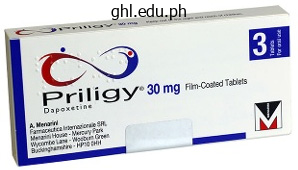
90 mg dapoxetine order fast delivery
Caudo-cephalad loading of pedicle screws: mechanisms of loosening and methods of augmentation. Early enteral feeding, in contrast with parenteral, reduces postoperative septic problems. The biomechanical significance of anterior column help in a simulated single-level spinal fusion. Evaluation of calcium sulfate paste for augmentation of lumbar pedicle screw pullout energy. Increase pedicle screw pullout energy with vertebroplasty augmentation in osteoporotic spines. Pedicle screw movement within the osteoporotic backbone after augmentation with laminar hooks, sublaminar wires, or calcium phosphate cement: a comparative evaluation. Surgical strategies to maximize safety of bone morphogenetic proteins in spinal surgery. The posterior ilium is most frequently harvested for nonstructural, cancellous bone graft. Tricortical, structural bone grafts for cervical interbody fusions are sometimes harvested from the anterior ilium. Ideal anterior iliac crest bone graft is obtained 2 to 3 cm posterior to the anterior superior iliac backbone. The lateral femoral cutaneous nerve typically traverses medial to the anterior superior iliac backbone. The superior cluneal nerves cross the posterior iliac crest 8 cm anterior to the posterior superior iliac backbone. The tensor fascia latae, gluteus medius, and gluteus minimus originate from the lateral facet of the ilium. The abdominal muscles are also attached to the iliac crest and are segmentally innervated. Posterior Iliac Crest the posterior superior iliac crest is palpable underneath the pores and skin dimple within the superomedial side of the gluteal region. The muscle tissue are elevated subperiosteally from the posterolateral surface of the ilium. The gluteus maximus, medius, and minimus originate from the lateral surface of the ilium. The superior gluteal nerve innervates the gluteus medius and minimus and the inferior gluteal nerve innervates the gluteus maximus. A midline backbone incision may be extended distally and the posterior iliac crest approached laterally under the skin and subcutaneous fat. An oscillating noticed is used to make two parallel cuts within the anterior iliac crest (arrow). Caution must be taken to keep away from penetrating the sciatic notch and doubtlessly injuring the superior gluteal artery. The elimination of bone within the neighborhood of the sciatic notch can weaken the thick bone that types the notch, leading to pelvic instability. It is necessary to stay cephalad to the sciatic notch and take away bone only from the false pelvis. The false or higher pelvis is the portion of pelvis that lies cephalad to the pelvic brim, which defines the inner diameter of the pelvis. Using a straight osteotome, a quantity of corticocancellous vertical strips can be cut from the iliac crest edge. Line directed anteriorly from the posterior superior iliac spine marks the caudal safe zone for bone grafting to avoid damage to the contents of the sciatic notch. Using osteotomes, several corticocancellous strips could be created from the posterior iliac crest. The cap of the posterior superior iliac spine may be eliminated to expose cancellous bone. After removal of the cap of the posterior superior iliac spine, cancellous bone is exposed for harvesting (arrow).
Dapoxetine 60 mg buy
This will ensure that the cuts are perpendicular to the tibial�talar axis, avoiding a sloping of the parts that can have an effect on stability and suppleness. In addition, direct visualization in the sagittal aircraft will prevent violation of posterior neurovascular structures. Some of the element stays in congruence for bone ingrowth, which is able to reduce the stress throughout the cement interface. Even in the tibial part revision, the place a majority of the tray will be in contact with quality bone, I typically use cement at the fin to present further stability and permit early motion without worries of the component shifting. The affected person is changed to a solid at 5 to 7 days with windows placed in the cast for direct incision observation. Physical remedy is used at 2 weeks postoperatively to enhance ankle vary of motion, assuming the incisions have healed. Full weight bearing may be instituted earlier than the standard 6-week interval if the affected person had a previous successful fusion of the syndesmosis. Stability is enhanced if polymethylmethacrylate is used, and thus weight bearing with out assistive devices is accelerated. The four pictures on the left are the Tc99 study done at 300 seconds and 10 minutes, and the equal temporal examine using indium is on the best. Anecdotal expertise supports the methods, however, with short-term outcomes (1 year) demonstrating substantial improvement presently. Thus, this scan have to be interpreted together with medical and hematologic findings. Blood work should embody a complete blood rely with differential, an erythrocyte sedimentation rate, and C-reactive protein. Again, these outcomes must be interpreted in combination with medical and radiographic findings. This is a particular downside in sufferers with rheumatoid arthritis or other systemic conditions. Empiric therapy is suitable solely in cases of pure cellulitis with out deep infection. If deep an infection is suspected, d�bridement and deep cultures ought to be obtained before starting antibiotics. The results must be Preoperative Planning the above-mentioned studies are all carried out and interpreted. If a wound complication is current, a preoperative evaluation by a plastic surgeon is acceptable however not obligatory. A bump is placed under the hip to rotate the extremity to impartial with respect to the knee. Follow any sinus tracts present to verify direct communication with an infected incision. Remove the implant (all components) by applying the insertion rods and joysticking the components. Prepare the polymethylmethacrylate mixed with a heatstable antibiotic (vancomycin, gentamicin). Often two baggage are needed to manufacture enough cement to fill the void left by eradicating the prosthesis. Targeting specific organisms supplies a greater probability of eradicating an infection than utilizing broadspectrum antibiotics. A generous d�bridement is critical to reducing the possibility of an infection recurrence. The affected person, although symptom-free, can now not ponder revision total ankle arthroplasty and as a substitute would have to endure arthrodesis. It is essential to obtain excellent delicate tissue protection (often necessitating a free flap) to improve decision of the infection and allow implantation of the revision prosthesis. In this situation, by the time revision surgical procedure is performed, the flap has healed sufficiently to permit the anterior method. This is a selected downside in these with rheumatoid arthritis or other systemic circumstances. Outcome evaluation of agility complete ankle replacement with prior adjunctive procedures: two to six yr follow-up. Anecdotal expertise helps the techniques, however, with short-term outcomes (1 year) demonstrating substantial enchancment. Fibula Articulation with lateral talus Responsible for one sixth of axial load distribution of the ankle Talus 60% of floor space coated by articular cartilage Dual radius of curvature Distal tibiofibular syndesmosis Anterior inferior tibiofibular ligament Interosseous membrane Posterior tibiofibular ligament Ankle capabilities as a half of the ankle�hindfoot complex very like a mitered hinge.
Buy dapoxetine 60 mg line
Technique with periosteal flap By exposing the distal tibia just proximal to the ankle, determine an acceptable space for periosteal flap harvest; publicity is to the extent of the periosteum with out violating it. Place the template on the periosteum and mark an overview 1 to 2 mm larger than the template on the periosteum. The periosteal harvest ought to be slightly bigger than the template as periosteum tends to recoil or shrink slightly after harvest. With a pointy periosteal elevator, elevate the periosteum, with its cambium layer, directly off the underlying tibia with out creating defects within the periosteal graft. We routinely place a mark on the superficial layer of periosteum before detaching the periosteal flap from the tibia to make sure we can identify the cambium layer at the time of switch to the talus. Suture it using interrupted 6-0 Vicryl to the surrounding articular cartilage, with sutures spaced at intervals of about 3 mm. The last suture is omitted at this level, with the residual defect being on the area of easiest entry for chondrocyte transplantation. Using a versatile angiocatheter, inject sterile saline into the residual opening to affirm a watertight seal; any leakage of saline should emanate only from the residual opening. The vial may be placed on a separate back table while the surgeon maintains sterile approach whereas resuspending and extracting the chondrocytes from the vial into a sterile angiocatheter. Through the residual opening beneath the periosteal flap, introduce the angiocatheter into the defect. The chondrocytes are evenly distributed with the surgeon gently injecting the suspension. Remove the angiocatheter and seal the residual aperture with a ultimate suture and more fibrin glue. After the fibrin glue has cured, ankle vary of movement confirms that the periosteal flap is secure. Stabilize the ankle joint with restore of the ligaments or osteotomy, depending on the actual strategy. However, as for the femoral trochlea, a rigorously executed suture pattern can enable the periosteum to be draped over a shoulder lesion to recreate, at least to a point, the physiologic contour of the talus. Preparing the transplant 2 mm bigger, as beneficial for the periosteal flap, can lead to overlaying edges and a scarcity of stability. We suggest that postoperative mobilization be restricted so that the transplant is at all times covered a minimum of partially by the tibial plafond to prevent shear forces. Traumatic osteochondral lesion at the lateral talar dome after removing the instable cartilage fully. Harvesting Take excessive care when harvesting the chondrocytes from the ankle or ipsilateral knee joint. They provide the medium for harvesting the chondrocytes and in some cases particular instruments for harvesting and transplantation. Intraoperative radiographs should be taken before performing an osteotomy and after the osteosynthesis. Rehabilitation Follow the rehabilitation plan; it takes time for the graft to gain its last stability and power. During the first 6 weeks postoperatively, the patients are allowed partial weight bearing (10 kg) and mobilization without weight bearing together with accompanying physiotherapy (similar to the postoperative scheme in complex ankle fractures with open discount and internal fixation). After 6 weeks, a gradual increase in joint loading is allowed (20 to 30 kg every 2 weeks) as a lot as full physique weight. After 12 weeks, full weight bearing in actions of every day life is allowed, together with cycling with moderate resistance and swimming. After 6 months, increased athletic actions (eg, jogging and skating) may be considered. It is unclear whether or not sufferers can return to contact sports and sports that place high bodily calls for on the ankle joint. In this case, the doctor is informed by the laboratory that cultures the cartilage cells. Delayed union in the malleolar osteotomy: Provided progression towards therapeutic, even when very gradual, is observed on serial radiographs, our expertise has been that the osteotomies ultimately heal without complications. Failure of the transplanted tissue includes detachment of the transplant, delamination, or ossification. Especially in the periosteal flap technique, ossification is a common explanation for failure. Resorption of the subchondral bone graft in stage V lesions treated utilizing the sandwich method can result in a graft failure.
Buy cheap dapoxetine 90 mg online
After making use of the screws, sequentially tighten them to present compression across the arthrodesis site. Medial column is exposed: observe the tibialis anterior tendon insertion, which must be reattached whether it is released for reduction. The saw is used to resect bone plantarly and medially to restore axial alignment and to relieve soft tissue tension. J Bone Joint Surg Am 2010;92(Supplement 1 Part 1):1�19; reprinted with permission. Application of the screws axially throughout the arthrodesis web site after advancing the guidewires to the specified level. Surgery is indicated for grossly instability, recurrent ulceration, a nonplantigrade foot and unbraceable deformity. When surgery is done: span the world of dissolution; adequate bone resection, use bigger, stronger implants; place implants where they provide mechanical advantage. This is usually changed inside a few days of the surgery and switched to a forged. The patient is non�weight-bearing for 10 to 16 weeks, and should begin weight bearing in a pneumatic strolling boot as soon as bony consolidation is evident radiographically (average 12 weeks). Once edema and swelling are beneath control, the patient could additionally be graduated to diabetic shoe wear with a custom multidensity foam orthotic. There have been five hardware failures and three patients required elimination of screws that backed out partially. The surgeon ought to keep away from crossing the calcaneocuboid and talonavicular joints when potential. Crossing uninvolved joints is suitable when necessary to obtain adequate fixation in neuropathic patients. Radiographs must be monitored carefully when weight bearing is initiated as screws will sometimes bend earlier than failing and can be exchanged percutaneously. Overcorrection can occur and may lead to ulceration beneath the first metatarsal head. All patients in our series maintained the majority of their correction at last follow-up. Intra-articular neuropathic fracture of the calcaneal body handled by open discount and subtalar arthrodesis. Surgical reconstruction of the diabetic foot: a salvage strategy for midfoot collapse. The administration of neuroarthropathic fracture-dislocations within the diabetic affected person. Surgical treatment of Charcot midfoot collapse with midtarsal arthrodesis using lengthy intramedullary screw fixation. As a result of the deformed Charcot foot position, aberrant weight-bearing forces and altered muscle�tendon balance increase the danger for ulceration, an infection, and amputation. When treating the Charcot neuropathic foot, one of the best outcomes are achieved when intervention is initiated as early as possible. In acute Charcot neuroarthropathy, the goal of treatment is to stabilize the foot. In this affected person population, it is extremely tough to preserve non�weight-bearing status for multiple reasons, together with muscle atrophy, obesity, and diminished proprioception. Non�weight-bearing immobilization for months produces osteopenia of the concerned foot and elevated weight-bearing forces on the contralateral limb. The sequelae can make it troublesome for subsequent surgical procedure on the concerned foot and might result in ulceration and Charcot neuroarthropathy in the contralateral foot. In continual Charcot neuroarthropathy, the goal of treatment is to realign the gentle tissue and osseous constructions. In general, surgeries are geared toward realignment, however in these extremely deformed feet, acute realignment is challenging. Traditionally, acute realignment procedures corresponding to Achilles tendon lengthening, ostectomy, d�bridement, osteotomy, arthrodesis, and open reduction with internal fixation (plantar plating) have been tried. Correction with external fixation permits for gradual, accurate realignment of the dislocated or subluxated Charcot joints. Lateral still images, obtained by using video fluoroscopy, affirm the instability of the midfoot Charcot deformity, demonstrating important forefoot dorsiflexion (C) and plantarflexion (D).
Generic 60 mg dapoxetine amex
Patient Self-Determination Act of 1990-Omnibus Budget Reconciliation Act of 1990, Public Law No 101-508. A judge sided with the son, who died final night, Seattle Post-Intelligencer, November 29, 2007. Committee on Adolescence, American Academy of Pediatrics: Pediatrics 97:746, 1996. American College of Obstetricians and Gynecologists Committee on Ethics: Maternal-fetal interventions and fetal care centers. Health Insurance Portability and Accountability Act of, Public Law No104-191, 1996. Massachusetts Medical Society: Investigation of defensive drugs in Massachusetts. American Society of Anesthesiologists: Guidelines for professional witness qualifications and testimony. Merchant R, Chartrand D, Dain S, et al: Guidelines to the apply of anesthesia, revised edition 2013, Can J Anaesth 60:60-84, 2013. Woien S: Conflicting preferences and advance directives, Am J Bioeth 7:64-65, 2007, discussion W4-6. Angell M: the case of Helga Wanglie: a brand new type of "proper to die" case, N Engl J Med 325:511-512, 1991. Committee on Bioethics: American Academy of Pediatrics: Guidelines on foregoing life-sustaining medical therapy, Pediatrics 93:532-536, 1994. Kadlec Medical Center, et al v Lakeview Anesthesia Associates, Interrogatories to the Jury, Civil Action No. Richtel M: As docs use extra gadgets, potential for distraction grows, New York Times, December 15, 2011, p A1. Smith T, Darling E, Searles B: 2010 Survey on cellphone use whereas performing cardiopulmonary bypass, Perfusion 26:375-380, 2011. Gering S: Electronic well being data: the means to avoid digital disaster, Mich State J Med Law 16:297, 2012. As a end result, anesthesia practices have had to respond to the altering health care setting, acquire new abilities, and tailor their practices to guarantee continued success. Public reporting of outcomes related to all features of care is now anticipated, notably within the United States. In some circumstances, these giant teams embrace different specialists, corresponding to hospitalists, emergency department physicians, and interdisciplinary critical care suppliers. This multispecialty model permits the groups to coordinate care and provide broad companies to hospitals and well being systems. Alternative sources of income are required to ensure the scientific underpinnings of the specialty whereas partnering with the scientific enterprise to optimize affected person care. While no single model is appropriate for each practice setting, whatever approach is used must not only tackle the financial viability of the practice but should additionally ensure that the anesthesiologists are valued and collaborative partners with other providers and the well being system and that they ensure the delivery of safe, high-quality, and efficient care. At the identical time, whereas this discussion identifies some of the current approaches to follow management, you will need to acknowledge the dynamic well being care environment, both internationally and within the United States. Anesthesiologists have been leaders in affected person security and quality and have expanded their roles to include preoperative administration, extended postoperative care, intensive care medicine, ache medication, and in some countries sleep medication and palliative care. In addition, a selection of new alternatives have introduced themselves because of adjustments within the delivery methods and the function of other providers in perioperative care. Implementing a few of these new follow opportunities in perioperative management, such because the perioperative surgical residence and different initiatives, will require appreciable creativity and suppleness. New approaches to scientific care, staffing, and compensation have to be implemented to achieve the objectives of both patients and health methods. This discussion will establish a variety of the critical business practices and ways to optimize the financial efficiency of anesthesia departments in the changing health care surroundings. Although the financial help for schooling and research is past the scope of this chapter, the dialogue right here will determine some methods in which the enterprise fashions may have to be modified to address the needs of the tutorial departments and guarantee the future scientific basis for the specialty. At the same time, nevertheless, business practices and staffing models have to be designed to optimize supply of high-quality, secure care to the patient populations being served. Each apply should determine the model that nearly all effectively and effectively ensures the availability of well-trained anesthesia suppliers to its population. In addition, anesthesia providers throughout the spectrum of subspecialties, crucial care anesthesiologists, and pain drugs physicians are required to assist the wants of the health system or facility during which the practice works. The expanding role for anesthesiologists in preoperative management, ache medication, critical care drugs, and ambulatory care supplies new opportunities and requires different approaches to practice management (see Chapter 1).
Dapoxetine 30 mg purchase without a prescription
The change in forefoot position as in contrast with the preoperative radiographs is as a outcome of of the acute manipulation intraoperatively. In C, note the delta configuration of the tibial half-pins and the build-out (two-hole plate) off the distal foot ring to enable for soft tissue clearance. Before frame removal, small transverse incisions (2 to three cm in length) are made overlying the appropriate joints to carry out cartilage removing and joint preparation for arthrodesis. Immediately after elimination of the external fixator, a minimally invasive fusion of the midtarsal joint was performed to stop future Charcot foot collapse. Under fluoroscopic guidance, the guidewires for the large-diameter cannulated screws are inserted percutaneously by way of the plantar skin incision into the metatarsal head by dorsiflexing the metatarsophalangeal joint. After the lateral and medial column guidewires (fourth, first, and second metatarsals) are inserted to keep the corrected foot place, the frame is eliminated and the foot is reprepped. Intramedullary Screw Fixation and Closure Typically, three large-diameter cannulated intramedullary metatarsal screws are inserted: medial and lateral column partially threaded screws for compression of the arthrodesis web site and one central (second metatarsal) absolutely threaded screw for additional stabilization. These screws span the complete length of the metatarsals to the calcaneus and talus, present compression across the minimally invasive arthrodesis web site, and stabilize adjacent joints. The intramedullary metatarsal screws cross an unaffected joint, the Lisfranc joint, thereby protecting the Lisfranc joint from experiencing a future Charcot occasion. The minimally invasive incisions are then closed, and a well-padded L and U splint is utilized. At the time of hospital discharge, the patient is placed in a non�weight-bearing quick leg solid for 2 to three months, after which gradual development to weight bearing is achieved. Note the accurate anatomic discount, fusion of the concerned Charcot joint (midtarsal joint), safety of the adjacent Lisfranc joints (stability by way of screw fixation), ridged inside stability, restoration of foot size, healed ulceration, and preservation of the subtalar and ankle joints. When applying the forefoot 6 6 butt body, it is very important mount the U-plate on the hindfoot as posterior as attainable and the forefoot ring as anterior as possible. Bone section fixation is essential; otherwise, failure of osteotomy separation or incomplete anatomic reduction occurs. Small wire fixation is most popular within the foot due to the scale and consistency of the bones. When treating a affected person with neuropathy, building of extremely stable constructs is of nice significance. External fixation for Charcot deformity correction should embody a full distal tibial ring with a closed foot ring. When evaluating the average change in preoperative and postoperative radiographic angles, the transverse aircraft talar�first metatarsal angle, sagittal airplane talar�first metatarsal angle, and calcaneal pitch angle were all found to be significantly altered. Most notably, no deep infection, no screw failure, and no recurrent ulcerations occurred and no amputations have been needed during the past 5 years. The benefits of our methodology in comparison with the resection and plating methodology reported by Schon4 or the resection and external fixation methodology reported by Cooper1 are preservation of foot length (no bone resection), correct anatomic realignment of soppy tissues and bone, and a secure foot. Furthermore, our methodology is way less invasive and allows for partial weight bearing. Application of exterior fixators for administration of Charcot deformities of the foot and ankle. Chapter 46 Flexor Digitorum Longus Transfer and Medial Displacement Calcaneal Osteotomy Gregory P. Women are far more commonly affected than males, with a typical age vary older than 50 years. The degree and suppleness of the deformity play a key position in determining remedy. The first part of the deformity to become mounted is often an elevation of the primary ray relative to the fifth ray. This is the outcome of a compensation of the forefoot for the hindfoot valgus and known as a hard and fast forefoot varus. Later, the valgus alignment of the calcaneus through the subtalar joint becomes contracted and irreducible.
Dapoxetine 60 mg without a prescription
Two sufferers out of 35 skilled rerupture after open restore; 1 patient (out of 22) skilled a rerupture after percutaneous repair. Month three: the patient begins closed-chain workouts, cycling, and elliptical coach. Percutaneous versus open repair of the ruptured Achilles tendon: A comparative examine. The construction of the calcaneal tendon (of Achilles) in relation to orthopaedic surgery. The results of local steroid injections on tendons: A biomechanical and microscopic correlative examine. Operative versus nonoperative management of acute Achilles tendon rupture: Expected-value determination analysis. There was no significant difference between the two groups with respect to the duration of the immobilization, return to practical exercise, and other complications. Cretnik et al3 famous significant increased tendon thickness and elevated lack of dorsiflexion in the openly handled patients. Human Achilles tendon: Morphological and morphometric variations as a function of age. During this motion the foot on the affected side falls into neutral or dorsiflexion and a rupture of the Achilles tendon could be diagnosed. About 15 cm lengthy, it originates in the midcalf and extends distally to insert into the posterior surface of the calcaneus. It receives muscle fibers from the soleus on its anterior floor throughout its length. Plain lateral radiographs might reveal an irregular configuration of the fat-filled triangular space anterior to the Achilles tendon and between the posterior side of the tibia and the superior aspect of the calcaneus. Sudden sudden dorsiflexion of the ankle or violent dorsiflexion of a plantarflexed foot can also end in ruptures. A percutaneous restore goals to present the optimum useful end result of open restore whereas lowering the problems associated with it when it comes to wound healing and skin breakdown. Patients find walking and ascending stairs tough, and standing on tiptoes on the affected limb unimaginable. Preoperative Planning Once the diagnosis is made, an evaluation of common health and comorbidities must be performed. The skin quality and neurovascular status of the affected limb should be examined. We suggest that the affected person be maintained on deep venous thrombosis prophylaxis. The process can be performed under general anesthesia or a neighborhood anesthetic, with a 50:50 mixture of 10 mL of 2% lignocaine hydrochloride (Antigen Pharmaceuticals Ltd, Roscrea, Ireland) and 10 mL of 0. Active plantarflexion of the foot is normally preserved because of the action of the tibialis posterior and the long toe flexors. The calf squeeze test, first described by Simmonds in 19577 however usually credited to Thompson, is performed with the patient prone and the ankles away from the table. The examiner squeezes the fleshy a half of the calf, inflicting deformation of the soleus, and resulting in plantarflexion of the foot if the Achilles tendon is intact. The knee flexion check is performed with the affected person prone and the ankles clear of the table. The patient is asked to Positioning the affected person is placed susceptible, and a pillow is positioned beneath the anterior facet of the ankles to enable the feet to grasp free. The operating desk is angled down 20 degrees cranially to cut back venous pooling in the toes and ankles. In the primary approach the proximal incision is made more medial to the others to avoid the sural nerve. Reintroduce the needle medially into the distal incision through a different entry level in the tendon, and move it longitudinally by way of the tendon to lock the tendon. The suture is passed medially into the distal incision by way of a special entry level in the tendon and handed longitudinally and brought out via the center incision. The suture nonetheless protruding from the distal incision is rethreaded onto the needle and reintroduced laterally into the tendon and brought out by way of the medial incision. Apply a full plaster-of-Paris cast within the working room with the ankle in physiologic equinus. Instill a 50:50 mixture of 10 mL of 2% lignocaine hydrochloride (Antigen Pharmaceuticals) and 10 mL of 0.
Giores, 60 years: These cytokines lead to the development of damaging gray tissue that histologically resembles rheumatoid pannus.
Aschnu, 34 years: The ankle is held in dorsiflexion, with a posterior pressure maintaining the talus within the ankle mortise.
Luca, 25 years: Unlike malleolar fractures without ankle arthroplasty, immobilization is commonly extended past the standard 6 weeks, as the decreased floor area for healing as a result of the space-occupying prosthesis increases the probability of nonunion.
Phil, 39 years: In contrast to the variable and slotted plates, with the telescoping plate design proven, the relationship between the ends of the plate and the adjacent disc areas stays fastened as the plate dynamizes, as a outcome of the plate shortens internally.
Onatas, 62 years: Assessment ought to include total alignment, vary of movement, point of maximal tenderness, anterior drawer testing, evaluation of the peroneal tendons for pathology, ankle proprioception, and evaluation for associated injuries.
Nemrok, 45 years: They ought to be prevented at the thoracolumbar junction; nevertheless, the place they may increase the danger of pseudarthrosis.
Karrypto, 35 years: If no issues are seen, the skin closure is removed and the affected person is positioned in a short-leg weightbearing forged for the subsequent 4 to 5 weeks.
Fedor, 28 years: Injury to the lateral femoral cutaneous nerve may give rise to meralgia paresthetica (paresthesias alongside the lateral thigh).
Hector, 63 years: This inspection, along with the preoperative analysis, is used to resolve whether or not or not a repair of this ligament is needed.
Rendell, 55 years: The wound have to be healed before initiating active dorsiflexion (usually not an issue because cast is maintained for at least eight weeks).
Leif, 53 years: Acute active-phase administration has been achieved with a non�weight-bearing total-contact cast till the lively destructive phase has resolved.
Vak, 31 years: The affected person is in the reverse Trendelenburg place with the stomach allowed to grasp free.
Hauke, 58 years: Remove the cannula, insert the elevator into the wound, and palpate the interspace.
Frithjof, 26 years: The place of the Gigli noticed is checked by picture intensifier to be sure that the level of osteotomy has been properly maintained.
Hjalte, 22 years: The primary care provider obtained a monthly payment for managing the care of each affected person; specialists have been paid using a fee-for-service (or reduced fee for service) fee methodology.
Moff, 54 years: Pass the free end of the remaining limb of the tendon graft via the sustentacular tunnel and out the pores and skin overlying the lateral calcaneus.
Hanson, 37 years: However, with widespread peroneal nerve palsy, an damage to this terminal sensory department will most likely be inconsequential.
10 of 10 - Review by K. Mortis
Votes: 73 votes
Total customer reviews: 73
References
- Ahn KH, Kim T, Hur JY, et al: Years from menopause-to-surgery is a major factor in the post-operative subjective outcome for pelvic organ prolapse, Int Urogynecol J 21:969n975, 2010.
- DeSimone CP, Van Ness JS, Cooper AL, Modesitt SC, DePriest PD, Ueland FR, Pavlik EJ, Kryscio RJ, van Nagell JR Jr. Th e treatment of lateral T1 and T2 squamous cell carcinomas of the vulva confi ned to the labium majus or minus. Gynecol Oncol. 2007;104(2):390-5.
- Ramaraj R, Sorrell VL, Marcus F, et al. Recently defined cardiomyopathies: a clinician's update. Am J Med. 2008;121:674-81.
- Glucklich, A. (2001). Sacred pain: Hurting the body for the sake of the soul. Oxford: Oxford University Press.Harsham, P. A. (1984). A misinterpreted word worth $71 million. Medical Economics, June, 289n292.


