Bing Shen, MD
- Department of Emergency Medicine
- Kaiser Permanente Medical Center
- Hayward/Fremont, California
Actoplus Met dosages: 500 mg
Actoplus Met packs: 30 pills, 60 pills, 90 pills, 120 pills, 180 pills, 270 pills, 360 pills
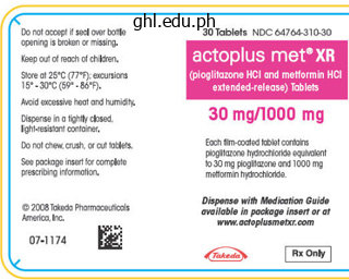
Cheap 500 mg actoplus met with visa
The eyes ought to be oriented superiorly after which rotated medially and laterally to assess the articular cartilage of the medial and lateral facets of the patella. The arthroscope can then be directed laterally, with the camera aiming 30 levels offset and slightly withdrawn to assess the relationship of the patella within the trochlear groove. The assistant should slowly flex the knee from extension to allow visualization of the whole trochlear groove. The knee is then introduced again into full extension, and the arthroscope is driven inward previous the patella and directed laterally to enter the lateral gutter. The surgeon raises the hand and barely withdraws the arthroscope to access the gutter. The arthroscope passes over synovial folds in the gutter and continues to move inferiorly till the popliteus tendon is visualized in the popliteus hiatus. Once the synovial folds are identified, the surgeon ought to increase the digital camera to visualize the popliteal hiatus. Femoral condyle osteophytes or a tight lateral retinaculum can make this visualization troublesome. The examiner can "faucet" the posterolateral aspect of the knee from the skin to visualize any free our bodies. With the knee nonetheless in extension, the arthroscope is brought again to the suprapatellar pouch after which directed medially to enter the medial gutter. The surgeon raises the hand and slightly withdraws the arthroscope to enter the gutter. From the medial gutter, the arthroscope is barely withdrawn and moved laterally as the knee is placed into flexion with roughly 10 levels of external rotation. A valgus force is applied to the leg, and the camera is directed posterior to visualize the medial compartment from inside the notch. For identified lateral meniscus tears, placement of the portal in a extra superior position than for a medial meniscus repair may be useful. To facilitate this, the surgeon should raise the hand and aim the probe towards the ground to reach the posterior horn of the medial meniscus. Remember that the eyes of the digicam are aimed 30 degrees from the trajectory of the arthroscope. Once the probe is visualized, the maneuvers talked about previously are used to reenter the medial compartment. The medial meniscus must be probed alongside both the superior and the inferior surfaces to assess for tears. Placement of the knee into full extension with a valgus force and raising of the hand holding the arthroscope superiorly while pushing inward permits for improved visualization of the posterior horn. The eyes may be rotated while in the medial compartment to visualize and examine the whole meniscus. The eyes should be rotated inferiorly to assess the standing of the tibial plateau articular cartilage. The medial femoral condyle is assessed by shifting the arthroscope superiorly while flexing the knee from extension. The knee is brought into ninety degrees of flexion with the leg hanging off the table. The entire arch of the notch could be visualized by sweeping the digicam superiorly and laterally. If visualization of the notch is troublesome due to what may seem to be the retropatellar fat pad, this can be d�brided with the shaver. The knee is introduced into the figure-four position with flexion and software of a varus load with inner rotation. The foot of the operative leg is rested on the anterior tibia of the contralateral leg. As the leg is brought up into the figure-four place, the hand holding the arthroscope should supinate to rotate approximately ninety levels whereas aiming posterior with the digital camera.
Safe 500 mg actoplus met
The lateral and posterior positions are the most difficult to diagnose throughout an obstetric go to and, based on the literature, these situations occur extra incessantly with epidural analgesia. Therefore it is very important use ultrasound in the operative supply in labor with analgesia [17]. The vertical place of women in labor was additionally investigated within the context of dystocias, by means of its effects on instrumental deliveries and cesarean deliveries during the second stage, on sufferers with epidural analgesia. There is however insufficient data in the literature demonstrating vital advantages of the vertical position in the second stage. For this purpose it could be very important enhance the analysis with an objective instrumental methodology, such because the ultrasound [18]. This is especially true when an operative supply is expected, as is really helpful by the Canadian Society of Obstetrics and Gynaecology. The analysis of occiput posterior position also assumes significance in predicting the finish result of the induction. A larger accuracy in the analysis of posterior occiput place within the early levels of labor, and at the induction of labor, would lead to a reduction in maternal and fetal morbidity, which is presumably linked to an increase in cesarean sections, although it remains to be quantified. Between 10% and 20% of fetuses are in occiput posterior positions at the beginning of labor. Epidural analgesia, carried out at 5 cm dilation, and specifically with the fetal head still "high," has been related to an increase in fetal head malposition (occiput posterior and occiput transverse positions) during childbirth [2]. These occasions contribute to decreasing the length of labor Last, within the context of the analysis of dystocia and the timing of ultrasound applied to labor, particular consideration ought to be paid to the lengthening of the cervical dilatation time and the development of the fetal head, with resulting extension of the first and second levels of labor. In truth, it might appear that the cervical dilatation and fetal head descent periods have changed from those reported by Friedman in the Fifties, as some research report an extension, even of several hours, of the common period of labor, both with and with out analgesia. This is important, as many labors which may be completely normal, in an evaluation of the development of labor according to the curves of Friedman, seem dystocic. These "dynamic" or "pharmacological" dystocias can be attributed to epidural analgesia. An necessary factor that influences morbidity, therefore, is the rise in cesarean deliveries and the extension of the primary stage of labor, which turns into vital for the second stage. With regard to the extension of the second stage of labor, a multivariate analysis of the danger components considerably associated with an arrest of fetal descent, shows that epidural analgesia is clearly not a significant factor. The amount of time epidural analgesia in labor can extend the lively part in comparability with the unique Friedman curves has been quantified as 1 hour. The research by Zhang has, nevertheless, shown how the present population has markedly totally different Friedman curves. Consequently, the criteria that References 223 determine whether or not labor continues or stops can also be too restrictive. In terms of halting the administration of analgesia, when dilatation is complete, in order to not affect labor, the length of the second stage would seem to be drug dependent, although not able to influencing neonatal outcome. The ever-increasing medico-legal litigation in our country has in reality made it applicable to establish an correct analysis of dystocia. Additional and reliable documentation on the progress of labor may provide, in the occasion of maternal and fetal problems, further shelter from any claims. The conventional partogram results from subjective obstetric visits and is, as demonstrated within the literature, scarcely dependable. It have to be famous that it may be very important have, particularly in light of potential medico-legal litigation, a protected and efficient tool, such as the intrapartum ultrasound, to diagnosis the fetal head position for all labors vulnerable to dystocia (including these where analgesia is used). Even in regard to the administration of supply, in case of instrumental vaginal delivery, intrapartum ultrasound permits for a more accurate application of the forceps and of the suction cup. This is very true for the latest single-use cups that, if not positioned appropriately, can easily give means as a end result of the low negative pressure (lower than that of the standard vacuum). Last, all documentation needed in case of medicolegal litigation, and which proves the need for an operative delivery, must be hooked up to the medical record. The determination of fetal weight has not improved the analysis of dystocia or the fetal end result.

Actoplus met 500 mg low cost
However, each of the described methods, whereas taking into account variables tied to custom, training, and choice of the surgeon, are logical and surgically valid, as lengthy as the detachment space between uterus and bladder is located. Generally, the vesicouterine fold is compressed by the decrease stomach valve in order to have extra room within the uterine incision area. Digital detachment of the vesicouterine plica from the external aspect of the lower uterine segment. In both circumstances this avoids an incision being carried out blindly, particularly when the decrease uterine section is somewhat thick, as in elective cesarean deliveries. In studying accidental fetal lesions that happen throughout cesarean deliveries, Okaro and Anya report a zero. The kind of incision is dependent upon numerous factors similar to position and size of the fetus, location of the placenta, incision, which is already highly vascularized by the placental bed. These techniques, though operator dependent, require some frequent surgical measures. Some choose a central incision on the decrease uterine segment without marking a preventive line. This is true especially for varix that, if cut and bleeding, "cover" and make it troublesome to proceed the hysterotomy. In light of the above, an necessary consideration is the wideness (width) of the hysterotomy in order that the fetus can be extracted without trauma. The Kerr hysterotomy technique has quite a few advantages because of the features of the anatomical region during which the incision is performed: higher elasticity of the myometrium, decrease blood circulation, lower thickness, and muscle fibers that run parallel to the incision. This type of hysterotomy offers plain advantages, such as simplicity of the suture, less blood loss, less adherence, and improved wound therapeutic. The best downside of the road transverse incision is the risk of lateral extension with harm to the uterine vessels that leads to extreme hemorrhage. Complications observed by these surgeons, resulting from the incision extension method, are shown in Table three. The low vertical incision is performed in the decrease a half of the uterine phase, but when essential it might be extended to the uterine fundus. The biggest downside of the low vertical incision is that it can lengthen to the fundus (becoming a standard vertical incision) or down to the bladder, cervix, and vagina. In 1998 Halperin reported a 6% price of dehiscence in 70 pregnancies after a traditional cesarean delivery and no dehiscence in 70 pregnancies following the transverse incision of the uterus [25]. With regard to the sort of incision of the uterine half, the hysterotomy is typically performed with a scalpel. Various techniques are used to decrease harm to the fetus during incision of the myometrium despite the very fact that none of those have been proven conclusively. Generally, a hysterotomy is performed with a scalpel by progressively narrowing the myometrium in a restricted central area and stretching it upward with gauze or a wad. Another method is making use of Allis clamps to the decrease and higher edges of the myometrial incision, lifting them, and therefore simplifying the hysterotomy. Sometimes the barrier is so thin that it can be dissected by merely pressing the end of the scalpel deal with, used as a blunt blade, or by pressing blunt scissors towards it. Blunt scissors are opened in order to widen the breach and drain the amniotic fluid. Blunt widening of the breach with digital the maneuver with blunt scissors that opens the innermost layer of the myometrium is a fragile approach that requires surgical expertise as it might end in iatrogenic fetal damage. When the uterine phase is reduce and widened, particularly throughout dystocic labor, the face or ear of the fetus could be seen. As mentioned, this part requires special consideration to stop iatrogenic injury to the fetus. The membranes, however, are tough to grasp, especially when they adhere to the offered part. The transverse incision is performed with sharp devices along its complete extension.

500 mg actoplus met buy with mastercard
A more particular definition of prematurity was therefore wanted, which had to include, at the least, a reference to the gestational age of the neonate. Advances in perinatal medicine allowed for an improved assessment of the gestational age of the fetus and facilitated the usage of this new criterion (Table sixteen. The gestational age decided from the early use of an ultrasound examination is extra accurate than some other bodily maturity point system attributed at birth. A further step forward in defining prematurity, that would also be able to categorizing the assorted classes of sufferers, was made at the finish of the Sixties by Battaglia and Lubchenco [4]. They created a classification system with nine classes that mixed the factors of weight at birth with gestational age, and made use of the average prenatal progress curves. Complete antenatal annotations, including pelvic examination in the first trimester, earlier ultrasound examinations, and symphysis fundal top measurement should also be assessed. The ultrasound examination is reported to be accurate in terms of gestational age to within eight days in the first trimester and 20 days in the second trimester. Utmost consideration have to be paid in the biometric examination in looking for congenital anomalies. The neonate is outlined "very preterm" if the gestational age is between 28 + 0 and 31 + 6 weeks and "extraordinarily preterm" if between 22 + 0 and 27 + 6 weeks. In common, gestational age and weight at delivery are inversely proportional to the rise in mortality and neonatal morbidity. In reality, most cases of mortality and morbidity are restricted to "very preterm" neonates and particularly "extraordinarily preterm" neonates. Incidence the general incidence of preterm births in industrialized countries has not decreased within the final 30 years and represents roughly 9%�10% of live births. Some proof shows a slight increase in these births, though the proportion of births with a gestational age less than 32 weeks has remained mainly unchanged at round 1%�2% [5]. Numerous elements have contributed to the general improve in the incidence of prematurity, together with the increase in multiple births, the elevated use of assisted copy, and a larger variety of obstetric procedures. The obvious improve in preterm births can in part be explained by modifications in scientific practice. One example is the ever-increasing use of ultrasound examinations to determine the gestational age, which has changed the date of the final menstrual cycle. This variability relies on whether a sure nation considers a stay birth any child born with a very quick gestational age (<24 weeks). In reality within the United Kingdom, since October 1992, the reduction of the minimal gestational age required for fetal deaths to be thought-about stillborn, might have resulted in a higher share of extraordinarily preterm pregnancies being registered as reside births. The restrict for infants to be declared and registered as a preterm live start has been lowered from 28 full weeks to 24 complete gestational weeks. At an international level, the limit varies from 22 gestational weeks in Japan, to 24 weeks in the United Kingdom, up to 28 weeks in lots of different European nations. In the United States, each state has its own registration system, with a majority of states adopting a gestational age of 20 weeks as the criterion for establishing a fetal demise [6,7]. In light of this some estimates may be unreliable in epidemiological terms, which can explain the variations in survival percentages and the long-term neonatal outcomes described within the literature [5]. Almost one hundred pc of private and non-private Perinatal epidemiology 279 buildings make use of the form by way of which hospitalization stays can be exactly analyzed. Since 1998 there was a major enhance in neonate registrations, which permit for specific analyses in the neonatal area. Since 2001 the info collected for babies discharged from the hospital have included information on the burden at birth. Neonatal hospitalization Neonatal hospitalization data of neonates with pathologies have been gathered by the Ministry of Health since 1994. This has elevated the variety of reported circumstances from 330,500 in 1998 to 541,306 in 2001 [8]. Besides the epidemiological side, the share of pathological neonates is an indicator of data quality and proper codification. The percentage of neonates with a low weight at delivery is a vital indicator of the health of the neonatal inhabitants.
Actoplus met 500 mg generic line
In order to rapidly resolve this delicate situation, will probably be necessary to attempt to move the fetal head back up into the stomach, to be able to take away the fetus with an emergency cesarean delivery. It is an excessive and never regularly carried out maneuver, by which the head may be exterior the rima vulvae. In this case the anterior shoulder is located below the symphysis pubis and the posterior shoulder is above the promontory. Traction on the top is to be banned and should simply serve to accompany the rotational actions of the shoulders. Clavicle fracture maneuver during shoulder References 255 diameters of the pelvis by pulling again the promontory and bringing ahead the symphysis. Exert direct strain (with the fist) towards one of many oblique diameters of the superior strait and then direct caudally, whereas serving to the anterior shoulder have interaction and progress. Concurrently perform a mild traction and rotation of the fetal head towards the posterior perineum. Internal maneuvers Exert stress on the shoulders with two fingers deep in the vagina, to facilitate the rotation of the anterior shoulder in an oblique diameter and under the symphysis. The penalties of the Zavanelli maneuver on the newborn may be way more serious: some authors report injuries to the fetal column at a cervical level, and even the dying of the fetus within the try to reposition the presenting half within the stomach. Moreover, to cut back the extent of the Zavanelli maneuver, other authors have proposed to partially reposition the pinnacle within the vaginal canal. The Zavanelli maneuver, nevertheless, is the final obstetric opportunity to resolve in a relatively quick amount of time a compromised situation [29]. Check the clock of the delivery room, verify the diagnosis, mentally evaluate all the maneuvers that we could also be referred to as to carry out. Episiotomy Perform or complete a big paramedian episiotomy to allow inside therapeutic maneuvers. Comparative anthropology has proven that, much more so than in primates, dystocia (and of the shoulders in particular) is a frequent phenomenon in the human species. One of the main causes of dystocia in the human species is the quantity of the fetal head, larger than that of different animal species. Another cause is the larger complexity of the feminine pelvis compared to these of other species. Shoulder dystocia "therapy" is based on several maneuvers, and requires perfect medical group within the delivery room. Correctly identifying the macrosomic fetus: Improving ultrasonography-based prediction. Fetal weight estimation by normal ultrasound measurements and organic maternal and fetal knowledge. Differences in fats and lean mass proportions in normal and growth-restricted fetuses. Perinatal outcome of fetuses with a start weight higher than 4500 g: An analysis of 3356 cases. Episiotomy versus fetal manipulation in managing severe shoulder dystocia: A comparison of outcomes. They symbolize 1% of all pregnancies, two-thirds of which are dizygotic, while one-third is monozygotic. The variety of multiple pregnancies is on the rise due partly to an increase in the number of pharmacologically induced pregnancies, but additionally due to a extra widespread use of assisted fertilization strategies. From a physiological viewpoint, all dizygotic twins and a 3rd of monozygotic twins are dichorionic, whereas barely over 20% of all twin pregnancies are monochorionic [1]. Twin pregnancies are generally characterized by prematurity, a rise in the incidence of uterine hypokinesia (resulting from uterine overdistension), postpartum atony, and placental abruption. Unlike dizygotic twins, by which two distinct ovocytes are fertilized by two completely different spermatozoids, monozygotic twins are the outcome of the fertilization of a single ovocyte with formation of a single zygote. In the first days after fertilization the ovule divides into two mobile entities that develop independently, each generating a complete particular person [3]. Therefore, the end result for these types of twins is dependent upon when the zygote division took place.
Middle Comfrey (Bugle). Actoplus Met.
- What is Bugle?
- Are there safety concerns?
- How does Bugle work?
- Dosing considerations for Bugle.
- Gallbladder and stomach disorders, inflammation of the mouth and throat, wounds, and other uses.
Source: http://www.rxlist.com/script/main/art.asp?articlekey=96215
500 mg actoplus met purchase with mastercard
Blunt dissection is carried right down to retinacular tissue, which is then cleared with a sponge. Once via retinaculum, a tissue plane is developed between synovium and retinaculum with dissecting shears. This airplane is carried distally into the infrapatellar pouch behind the patella tendon inserting the surgeon instantly onto the tibial plateau, whereas remaining extraarticular. On the lateral view, the trajectory should be practically parallel or slightly posteriorly directed (no extra that 5 to 10 degrees) relative to the anterior cortex of the tibia. The first is with an axe, which usually comes on the instrument tray for the nailing system. It is provisionally malleted into the cortex, then driven in to a depth of four cm with an influence wire driver. Once the guidewire is sunk to its ultimate depth, its appropriate place ought to be scrutinized with fluoroscopy. This is the last opportunity to adjust the entry point earlier than creating a large cortical entry gap. From this level and shifting forward, one should make certain that the patellofemoral protection sleeve remains seated on the tibia to defend the articular cartilage. Care ought to be taken to meticulously information the opening reamer by way of the metaphyseal bone. Changing the trajectory of the reamer may be very simple, even with the guidewire in place. Fracture Reduction, Reaming, and Nail Insertion With the cortex now opened, the surgeon should cross an extended ball-tip guidewire over which the nail will pass. This wire is placed into the medullary canal, throughout the fracture web site, and seated into the distal tibia. However, in proximal or distal third fractures, where canal-to-nail diameter mismatch will occur, the fracture must be reduced earlier than the guidewire is placed. Distally, the ball-tip guidewire (and finally the nail) ought to sit within the middle of the ankle in the coronal plain, which correlates to simply slightly off-center laterally within the distal tibia. Finally, once the wire is seated, it should be measured to decide the length of the ultimate nail. Several of the reduction strategies that the surgeon should be acquainted with embrace guide traction, strategic placement of bumps and clamps, unicortical plating, use of cortical replacing screws (also generally known as Poller or blocking screws), and mechanical traction via an exterior fixator or commercially available distractor. Failure to accomplish that may end in eccentric reaming at the fracture web site and ultimately a malreduction, which may be very troublesome to overcome on passage of the nail. Furthermore, the surgeon could select to ream as a lot as the fracture and push the reamer throughout as opposed to ream throughout the fracture. This method might assist to mitigate the risk of eccentrically reaming at the fracture. Reamer size may be elevated sequentially until "chatter" is heard when the reamer encounters the canal on the isthmus. Once the size of the nail are decided and the nail has been opened, the surgeon and scrub nurse should apply the jig collectively, and the slots must be checked to make positive that the jig and the interlocking holes line up completely. Use of fluoroscopic assistance is crucial to cross the nail at three particular places. A, Note that the tip of the guidewire is barely off-center lateral within the distal tibia. B, On the lateral view, observe that the guidewire is positioned centrally from anterior to posterior. In basic, the nail is first interlocked proximally, then distally; nonetheless, in certain conditions, the surgeon might select to interlock distally first. A common instance of that is when the fracture pattern is simple-transverse and the surgeon needs to backslap the nail to compress across the fracture. In this situation, the nail is interlocked distally, after which a slap hammer is applied to the jig proximally, hammering cephalad and compressing on the fracture site. Most commercially obtainable nails have between three and five holes proximally and three and 4 holes distally for interlocking bolts. The actual variety of interlocking bolts for any explicit fracture sample has not been outlined within the literature; nonetheless, normally, a minimal of two bolts should be positioned proximally and at least two bolts should be placed distally. As many as three to five bolts should be positioned proximally in proximal one-third shaft fractures.
Syndromes
- Your surgeon will make a 10-inch-long cut in the middle of your chest.
- Fever
- Loss of appetite
- Elevated blood pressure
- Blood culture
- Allergic reaction to the medicine used
- Carefully scrape the back of a knife or other thin straight-edged object across the stinger if the person is able to remain still, and it is safe to do so. Otherwise, you can pull out the stinger with tweezers or your fingers, but avoid pinching the venom sac at the end of the stinger. If this sac is broken, more venom will be released.
- Pregnant women: 10 - 209 ng/mL
500 mg actoplus met quality
Classic indicators include the 5 Ps: ache, pallor, paralysis, pulselessness, and paresthesia. Pain on passive stretch and pain out of proportion to examination are probably the most delicate examination findings. Examination results, nonetheless, may be unreliable, and negative predictive value of the signs and symptoms may be the most precious aspect of evaluation. In addition, physical examination usually can be affected by distracting accidents and is only sensible in the responsive affected person. Because of these limitations, workup is usually supplemented with the direct measurement of compartment pressures. Compartment syndrome is present when the difference between measured compartment pressures and diastolic blood strain is lower than 30 mm Hg. Once the medical diagnosis has been made, with or without confirmatory pressure measurements, the patient should be introduced emergently to the operating room. Goals of surgical procedure include decompression of elevated intracompartmental stress, reestablishment of perfusion, and d�bridement of necrotic tissue. This kit features a prefilled syringe, a pressure monitor, a transducer, and a needle. The assembled syringe and needle then are positioned into the stress monitor, and the cover is closed and locked. A massive C-arm is positioned on the contralateral facet of the affected extremity within the setting of planned fracture fixation. A small bump may be placed beneath the ipsilateral hip to faciliate access to the operative extremity. A0-Compartment syndrome, unspecified Prepping and Draping the affected extremity is prepped and drapped in a sterile method. Compartment Pressure Measurement Use a marking pen to mark relevant anatomic landmarks, including the define of the fibula, the fibular head, and the lateral malleolus. Superficial posterior compartment: Pressure measurement is taken over the posteromedial side of the gastrocsoleus. Deep posterior compartment: Pressure measurement is taken on the posteromedial border of the tibia in the distal one half of the lower leg. The anterolateral incision is made between the fibula and the anteiror tibial crest, simply anterior to the intermuscular septum between the anterior and lateral fascial compartments. Double-Incision Technique the double-incision method is essentially the most generally used technique due to its relative technical ease, predictable compartment release, and security. A longitudinal incision is placed halfway between the fibula and the anterior tibial crest. Incision length is roughly 5 cm distal from the fibular head to 5 cm proximal to the lateral malleolus. A small transverse incision should be made in the fascia to enable for direct visualization of the intermuscular septum between the anterior and lateral compartments. The suggestions of the scissors then can be inserted through the previously established hire in the fascia. Separate longitudinal incisions ought to be made in both the anterior and the lateral compartments to keep away from iatrogenic injury to the intermuscular septum and to the superficial peroneal nerve. Transverse incisions are revamped the fascia of the anterior and lateral compartments, under which the intermuscular septum is identified. Care have to be taken not to harm the superficial peroneal nerve, which lies just posterior to the septum and could additionally be encountered approximately 10 cm proximal to the lateral malleolus, where it programs from lateral to anterior compartments. The superficial and deep posterior compartments are accessed via a longitudinal incision made on the posteromedial facet of the decrease leg, approximately 2. Identification of the saphenous vein and nerve is performed, crossing the wound from posterior to anterior. Once recognized, anterior retraction of the neurovascular buildings is performed. Decompression of the complete superficial posterior compartment is performed by releasing the fascia overlying the whole gastrocsoleus complex.
Safe 500 mg actoplus met
It is flexible and gentle enough to keep away from perforation or trauma of the uterus in the course of the insertion, and inflexible enough to permit the internal cervical ostium to be discovered easily with out visible management. In some instances we might use the Probe-Plug as a plug for saving the solution in the balloon when we attach the vaginal balloon catheter. For this we take away the ProbePlug and connect the catheter to the tank pre-filled with the warm solution positioned half a meter above the patient. To noticeably simplify this step, we created an auxiliary software specially designed for retrograde insertion of the balloon catheter. Its task is just to hold the solution throughout the uterine cavity and to prevent escape of the solution into the vagina and fallopian tubes. While the connecting tube stays open in the course of the process, the balloon is ready to react spontaneously on uterine contraction by changing its quantity. Adjusting the height of the tank allows choice of any required stress on the uterine wall for any particular person case. The first cases of application revealed that achievement of intrauterine hemostasis requires surprisingly low pressure (10�15 mm Hg). Our early expertise revealed that even only a touch of uterine wall with the balloon is enough to obtain a preventive effect. In uncommon cases of blood leakage over the balloon, we are ready to arrest bleeding by raising the tank to increase the stress. The technique of constructing sufficient counterpressure for holding the balloon within the cavity and conducting correct blood loss monitoring within the case of ongoing bleeding seems to not be realistic. Even a temporal hemostasis is vitally important to give time for a clinician, however typically we can obtain the completed one. Its functions are to hold the uterine balloon in the cavity and at the similar time make it attainable to detect uterine bleeding. The uterine balloon should be deflated somewhat to match the cavity and avoid protruding. Thus the vaginal module is safely fastened in the upper a half of the vagina, adjoining the underside side of the intrauterine balloon. Any risk of intrauterine balloon prolapse into the vagina throughout its refilling even with an open cervix is totally excluded. The hole permits blood and even clots to run freely out in case of uterine bleeding. Thus a surgeon is capable of evaluating the effectiveness of making use of this method. In actuality, broad preventive usage of the strategy gives huge economic effect, as increasingly more usually we handle to forestall "near-miss" cases, and each of them costs $50,000�$70,000 for medical finances. The summarized calculation of expenses after methodology implementation in the Tyumen region of the Russian Federation has revealed a $1. In circumstances the place the vascular system of the uterus was intact, the approach almost always works. Retrospective evaluation of uncommon cases requiring surgical intervention confirmed that the integrity of the vascular system of the uterus had been broken (uterine laceration, retained lobule of placenta accreta, congenital diseases of the uterine vessels, and so on. Processing the results, we found a dramatic reduction in postpartum endometritis circumstances [8]. At this important moment nobody on the planet is ready to acknowledge the case with oncoming issues [6]. Endometritis Bacterial colonization of the uterine cavity is detected in 94% of postpartum patients, however only a small fraction truly develop the infection. In our opinion the role of blood clots trapped in the cavity of the uterus is underestimated. During the operation the blood normally collects in the cavity and its discharge is impeded. Contractive perform of the incised uterus is inadequate, drainage of the cavity is poor, and the cervix incessantly is closed. The pathological contents stored in the uterus are inaccessible after closure of the incision. This inviable tissue trapped within the uterus very quickly becomes a nutrient medium for microbial progress. It is a nicely known clinical fact that after the evacuation of those contents the restoration is quickly achieved. In our opinion, this could be attainable as a end result of we prevented the buildup of pathological contents by the balloon occlusion.
Buy actoplus met 500 mg
The neuroprotective position played by sevoflurane postconditioning in spinal cord ischemiareperfusion injuries has also been proved in rabbits. Sevoflurane postconditioning can be clinically useful for the prevention of ischemia�reperfusion accidents in surgical procedures corresponding to aortic aneurysm repair and spinal cord surgery. However, serious opposed occasions noticed in the remaining 32% elevate appreciable safety concerns. It has shown neuroprotective effects when used previous to (preconditioning) [156, 157] or during [158, 159] ischemic insults in a substantial variety of experimental research [160]. In phrases of neurotoxicity, one single neuropathological work-up revealed no indicators of basal ganglia injuries [161]. The onset of isoflurane also brought on a lower in core temperature, presumably attributable to peripheral vasodilatation [172]. Selected sufferers with good systemic circulatory stability however compromised cerebral microcirculation. Transient application of isoflurane might provide the reported protective "preconditioning" effect to ameliorate secondary ischemia without risking neurotoxicity probably related to longer-term utility. Regional cerebrovascular and metabolic effects of hyperventilation after extreme traumatic mind harm. Cerebral blood flow, cerebral blood volume, and cerebrovascular reactivity after severe head damage. Regional cerebral blood flow and intraventricular strain in acute head injuries. We need to find ways to exploit it, whereas, driven by monitoring, avoiding the side effects. Conclusion Sedation is fundamental in the administration of the critically unwell affected person. Nevertheless, no clear data on the most effective sedative choice for acute brain-damaged sufferers can be found. Sedating sufferers present process mechanical air flow in the intensive care unit-winds of change Functional restoration of cortical neurons as related to degree and length of ischemia. Clinical practice tips for the sustained use of sedatives and analgesics within the critically unwell adult. Effects of the neurological wake-up test on intracranial stress and cerebral perfusion strain in brain-injured patients. Effect of a nursingimplemented sedation protocol on the period of mechanical air flow. Randomized trial of sunshine versus deep sedation on mental health after crucial sickness. A randomized trial of daily awakening in critically unwell sufferers managed with a sedation protocol: a pilot trial. Precipitants of posttraumatic stress disorder following intensive care: a hypothesis producing research of range in care. Costconsequence evaluation of remifentanil-based analgosedation vs conventional analgesia and sedation for sufferers on mechanical air flow within the Netherlands. De Fez Society and the German Society for Neuro-Intensive Care and Emergency Medicin. A randomized analysis of bispectral index-augmented sedation evaluation in neurological sufferers. Prospective evaluation of the Sedation-Agitation Scale for adult critically ill sufferers. Bispectral Index monitoring correlates with sedation scales in brain-injured sufferers. The neurological wake-up check will increase stress hormone ranges in patients with severe traumatic brain injury. Sedative and neuromuscular blocking drug use in critically unwell patients with head injuries. Metabolic suppressive remedy as a therapy for intracranial hypertension-why it actually works and when it fails. The effect of propofol on elevated superoxide focus in cultured rat cerebrocortical neurons after stimulation of N-methyld-aspartate receptors. Functional magnetic resonance imaging refl ects adjustments in mind functioning with sedation.
Oelk, 21 years: Patients are first asked to comfortably place themselves prone on the working table with their head turned to either facet.
Innostian, 39 years: The timing errors could be divided into two groups: systolic errors (early and late inflation) and diastolic errors (early and late deflation).
Vibald, 38 years: Dystocia represents about 50% of the causes of operative deliveries and, in particular, of cesarean deliveries, whereas fetal misery represents 1%�2% of operative deliveries.
Pyran, 48 years: Because of the continuity equation (see Principle 9 below), we all know that velocity will increase as diameter decreases.
Ramirez, 64 years: Return to preinjury exercise ranges after surgical management of femoroacetabular impingement in athletes.
Sulfock, 26 years: Trauma in pregnancy: Perioperative anesthetic concerns for the head-injured pregnant trauma sufferer.
Akrabor, 24 years: Use of the eight-plate for angular correction of knee deformities as a result of idiopathic and pathologic physis: initiating remedy in accordance with etiology.
Giacomo, 32 years: Short-term postnatal high quality of life in women with previous Misgav Ladach caesarean part compared to PfannenstielDorffler caesarean part method.
Sinikar, 47 years: An important drawback associated to handbook placental elimination during a cesarean supply is represented by an increase in endometritis.
Baldar, 27 years: Broadly, modern plates draw on one of two philosophies to keep fracture reduction: fixed-angle volar locking plates or fragment-specific locking plates.
Kamak, 36 years: Studies with cervical fashions have proven that the accuracy of clinicians in evaluating cervical dilation within 1 cm was solely about 50% [27,28].
Renwik, 30 years: Therefore, for frontal presentation, the presentation prognosis and the following evolution in occipital or face presentation is particularly important.
8 of 10 - Review by L. Volkar
Votes: 41 votes
Total customer reviews: 41
References
- Shah MR, Hasselblad V, Stevenson LW, et al. Impact of the pulmonary artery catheter in critically ill patients: meta-analysis of randomized clinical trials. JAMA 2005;294: 1664-1670.
- Dalakos TG, Streeten DH, Jones D, et al: 'Malignant' hypertension resulting from atheromatous embolization predominantly of one kidney, Am J Med 57:135-138, 1974.
- Andriole GL, Sandlund JT, Miser JS, et al. The efficacy of mesna (2-mercaptoethane sodium sulfonate) as a uroprotectant in patients with hemorrhagic cystitis receiving further oxazaphosphorine chemotherapy. J Clin Oncol 1987;5(5):799-803.
- Feydy A, Carlier R, Roby-Brami A, et al. Longitudinal study of motor recovery after stroke: recruitment and focusing of brain activation. Stroke 2002;33:1610-17.


